COMMUNITY DIGEST
3 featured cases and 8 quick consultations from the EndoCollab WhatsApp community between July and September 2026. Exposed sutures two years after endoscopic sleeve gastroplasty, two polypoid lesions beside the ampulla on a pre-bariatric endoscopy, and an ascending-colon lesion taken out en bloc. Quick consultations cover prolapse-related rectal change, lymphocytic colitis behind bland mucosa, a duodenal nodule, and several decisions the group argued through.
In this issue
- Exposed Sutures Two Years after Endoscopic Sleeve Gastroplasty
- Polypoid Lesions beside the Ampulla on a Pre-Bariatric Endoscopy
- Ascending-Colon ESD: Paris IIa plus IIc, NICE 2B
- Quick cases: Four-day colitis in psoriasis, biologic held; Bland rectal mucosa, lymphocytic colitis on random biopsies; Pale nodular lesion in the horizontal duodenum; Metallic object in a dilated distal ileum; Polypoid rectal lesion with known prolapse; Full-thickness clip: the click was not deployment; Jejunal erosions, histology still open; Erythema in the diverted limb
- Community notes: Standalone acetic acid mix named for flat lesions and pit bo…; An isolated esophageal venectasia is an observation. Leave i…
Featured Cases
1. Exposed Sutures Two Years after Endoscopic Sleeve Gastroplasty
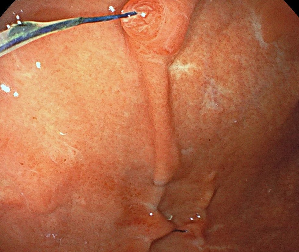
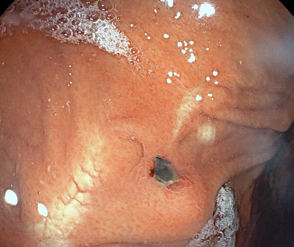
A woman in her late thirties had an endoscopic sleeve gastroplasty at another hospital two years earlier. The weight stayed down. For one to two months she had new left-upper-quadrant pain, worse with eating. The abdomen was soft and the blood tests were normal.
Gastroscopy showed exposed sutures, inflammation around the sutures, and partial mucosal bridging. There was no torsion and no stenosis. It was the first gastric sleeve this endoscopist had inspected.
The group treated loose sutures and local inflammation as expected this far out. Fibrosis and slower emptying can hold the weight loss even when sutures are visible. New focal pain still needs cross-sectional imaging for another cause. Cutting the sutures is not the default next step.
Clinical Pearl: Exposed sutures and peri-suture inflammation two years after ESG are expected. They do not show that the sleeve has failed. Image new focal pain before anyone cuts suture.
2. Polypoid Lesions beside the Ampulla on a Pre-Bariatric Endoscopy
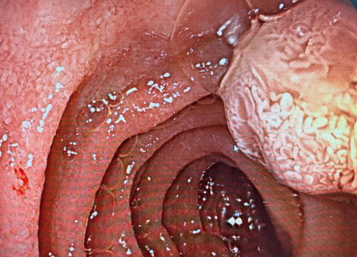
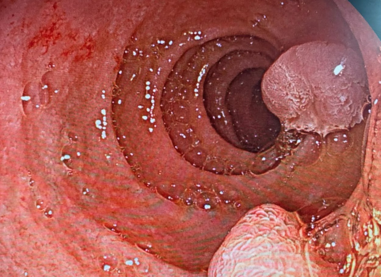
Pre-bariatric endoscopy in a 20-year-old showed two polypoid lesions in the second duodenum. In the room they were called a prominent major papilla and minor papilla, because the distal lesion seemed to have an orifice.
Still review made that label uncertain. One read was inflammatory polyps, and the ask was to biopsy. Another read said this was not the ampulla and that the distal lesion might be adenomatous. The plan was to bring her back, biopsy, and use a side-viewing duodenoscope if the forward view stayed unclear.
A normal-papilla label is the wrong clearance for bariatric surgery when the stills look polypoid.
Clinical Pearl: Polypoid lesions beside the ampulla need a biopsy. If the forward view is unclear, use a side-viewing duodenoscope before anyone calls them normal papillae.
3. Ascending-Colon ESD: Paris IIa plus IIc, NICE 2B
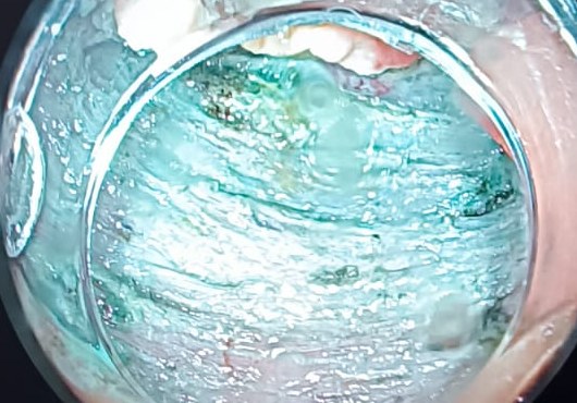
An ascending-colon Paris IIa plus IIc lesion, read as NICE type 2B, was removed en bloc by ESD in about an hour with a 3 mm FlushKnife. The operator reported a clear submucosal plane and no major complication.
The still is that plane during the resection: blue-green injectate, a lifted mucosal flap, and the dissected submucosa. The group did not debate who should attempt colonic ESD. The limit worth keeping is the one in the note: en bloc resection when access lets the plane stay visible.
Clinical Pearl: A Paris IIa plus IIc, NICE 2B colon lesion can still be an en bloc ESD when the submucosal plane stays clear. Keep a still of that plane, not only a finished specimen.
Quick Consultations
Colitis · Four-day colitis in psoriasis, biologic held
A middle-aged man with psoriasis and a history suggesting axial spondyloarthritis had four days of rectal bleeding and pain. Differentials named in the thread were enteropathic colitis, ulcerative colitis, and Crohn disease. Biopsies showed no chronicity. The advice was to hold a biologic, send cultures including C. diff, ask for CMV stains, and start with a topical 5-ASA while those results come back.
Colon · Bland rectal mucosa, lymphocytic colitis on random biopsies
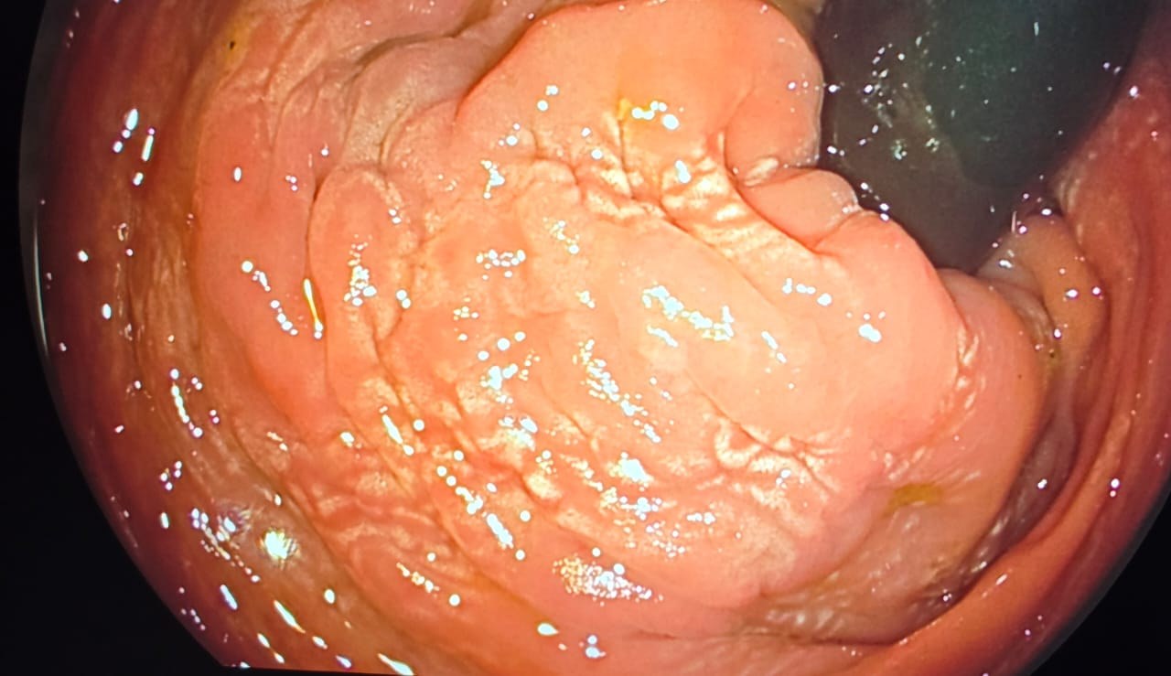
Diarrhea workup. Rectal mucosa looked nearly normal, with a whitish pattern read at first as nonspecific lymphangiectasia. Random colonic biopsies, rectum included in the same jar, came back as lymphocytic colitis. Keep the random biopsies when the rectum looks bland.
Duodenum · Pale nodular lesion in the horizontal duodenum
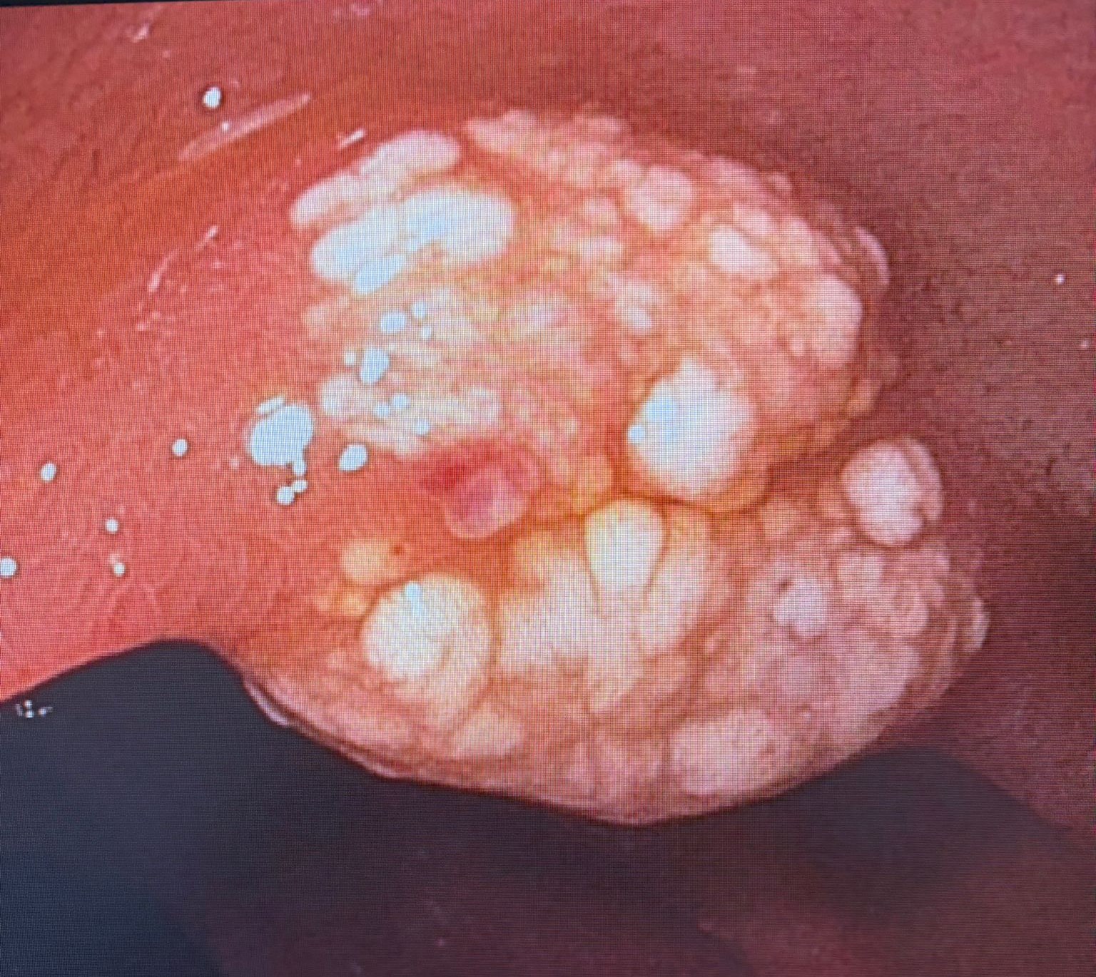
A pale, nodular, mammillated lesion in the horizontal duodenum. Reads offered: Brunner gland hyperplasia, especially after peptic injury, plus xanthoma, lipoma, and lymphangiectasia or lymphangioma. Biopsies were taken. Wait for histology before you name it.
Foreign body · Metallic object in a dilated distal ileum
CT in an older patient described a metallic foreign body in a dilated distal ileum and raised concern for impending perforation. Do not give a full bowel prep and push a colonoscopy through that. If endoscopic retrieval is chosen, a cap and fluoroscopy help after the ileum is intubated.
Rectum · Polypoid rectal lesion with known prolapse
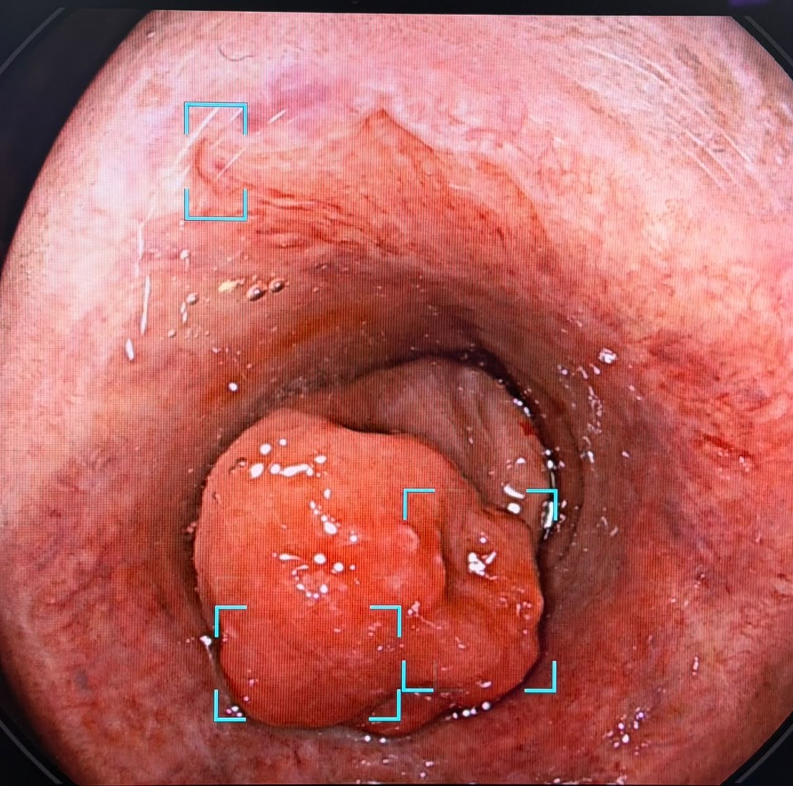
An 83-year-old woman with known uterovaginal prolapse presented with rectal bleeding. Colonoscopy showed a polypoid rectal lesion, and the question was solitary rectal ulcer syndrome. The group read was SRUS. With that history, biopsy the lesion and treat the prolapse. Do not resect it as an ordinary adenoma on the first look. The blue boxes on the still are the processor detection overlay, not a marked margin.
Resection · Full-thickness clip: the click was not deployment
First full-thickness resection, a descending-colon lesion, with severe sigmoid diverticulosis in the way. The operator heard the click, was not sure the clip had actually deployed, then cut. The resection was not full thickness, an over-the-scope clip closed the site, and the patient did well. After the grasper came out, the release cable had gone slack and needed the hand wheel to restore tension. Confirm the clip is on the wall and the cable is tight before you close the snare. One practical suggestion was to have the device representative in the room for the next case.
Small bowel · Jejunal erosions, histology still open
Follow-up only, on the multifocal jejunal lesions from the last digest. The added point was a pathology review of the mucosa. Chronic changes would favor inflammatory bowel disease, and CMV can ride along. The capsule stills alone did not settle drug injury versus something else.
Stoma · Erythema in the diverted limb
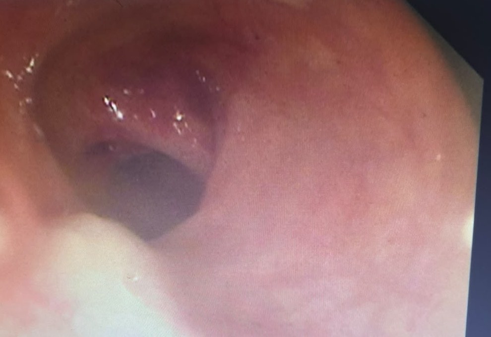
A 75-year-old woman, two months after resection of gangrenous transverse colon, now with a prolapsing colostomy. The antegrade limb from the stoma to the cecum looked normal. The diverted limb, rectum toward the splenic flexure, was erythematous with an altered vascular pattern. The group read was diversion colitis. The still is a screen photo and it is soft. Treat it as a pattern match, and wait on the biopsies.
Community Notes
- Chromoendoscopy: Standalone acetic acid mix named for flat lesions and pit borders: 75 ml of sterile 5% acetic acid plus 175 ml of sterile water or saline (250 ml, about 1.5%). Spray through a catheter and wait 10 to 15 seconds. Confirm the stock concentration before you mix it. Do not substitute pantry vinegar.
- Esophagus: An isolated esophageal venectasia is an observation. Leave it alone. Therapy is for varices.
What’s Next
Every case in this digest came from real conversations in the EndoCollab private WhatsApp community, 650+ practicing endoscopists discussing cases daily from every continent. Lifetime members get permanent access to the group, plus the full EndoCollab case library.