COMMUNITY DIGEST
3 featured cases and 4 quick consultations from the EndoCollab WhatsApp community between September 23 – October 8, 2026. Inflammatory pseudopolyps after Crohn colitis healed, low-grade dysplasia inside a postoperative stricture the scope could not pass after two dilations, and an ERCP where a loop in the stomach made the papilla look like third-part duodenum. Quick consultations cover carpeted antral polyps, delayed bleeding from banding ulcers, common duct stones with a clear gallbladder, and a cyst after hydatid surgery.
In this issue
- Inflammatory Pseudopolyps after Crohn Colitis Healed
- Low-Grade Dysplasia inside a Postoperative Crohn Stricture
- ERCP: A Gastric Loop Made the Papilla Look Like Third-Part Duodenum
- Quick cases: Common duct stones, clear gallbladder, no surgery wanted; Anechoic cyst after hydatid surgery; Carpeted antral polyps with a normal colon and duodenum; Delayed bleeding from banding ulcers, INR above 2.5, platelets 35,000
Featured Cases
1. Inflammatory Pseudopolyps after Crohn Colitis Healed
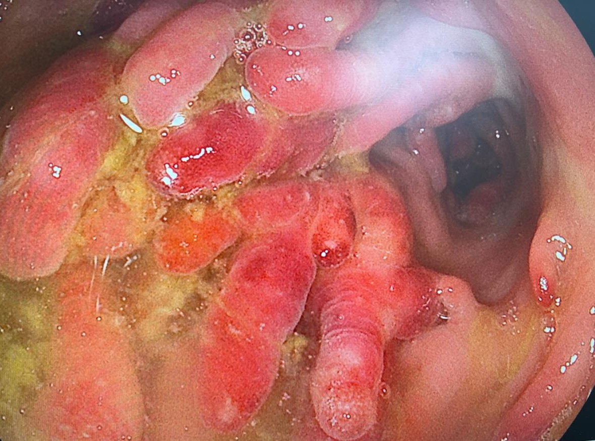
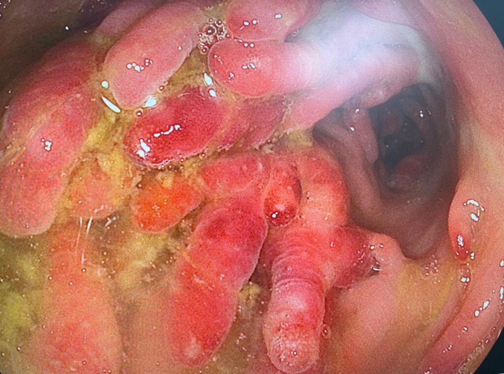
A patient with ileocolonic Crohn disease once had a fistula and very deep ulcers in the sigmoid and distal left colon. On infliximab every four weeks, the ileal ulcer and the anal fistula healed, and he felt well. Colonoscopy now showed a field of inflammatory polyps from 32 to 37 cm. The two largest were removed. The longest measured 2.6 cm. Histology was pending.
The group split on the rest. One view: pseudopolyps form when inflammation settles, so they show that the treatment worked. They are not a reason to raise the dose. Practice guidance cited in the thread favors sampling representative or suspicious pseudopolyps rather than removing all of them. In the thread, removal was reserved for polyps that are symptomatic, look suspicious, or block surveillance. One member also removes them when they bleed. Another member said the surrounding mucosa is the bigger worry, because it marks severe past inflammation and a higher cancer risk.
When removal is chosen, one member's method is to inject epinephrine first, take the polyp from its root with a hot snare, and use a little coagulation before a pure or blended cut. He avoids leaving a clip on a completed cut, because a clip may make the polyp come back. If the cut bleeds, his order was clip, then injection, then Hemospray, then APC. They bleed easily. In one of his patients, clearing them took about six sessions. One polyp held high-grade dysplasia and a few held low-grade dysplasia. In another patient, with ulcerative colitis, one odd-looking pseudopolyp held cancer. He said that was not a common finding.
On the drug: raising the dose is unlikely to shrink the polyps. Check a trough and adjust the infusions to the target for healed fistulizing disease. Fixed-dose subcutaneous infliximab, 120 mg every two weeks, is an alternative that needs no level checks.
Clinical Pearl: Pseudopolyps after Crohn colitis heals usually mean the treatment worked. Sample the suspicious ones. Remove those that bleed, cause symptoms, look suspicious, or block surveillance. A higher biologic dose is unlikely to shrink them.
2. Low-Grade Dysplasia inside a Postoperative Crohn Stricture
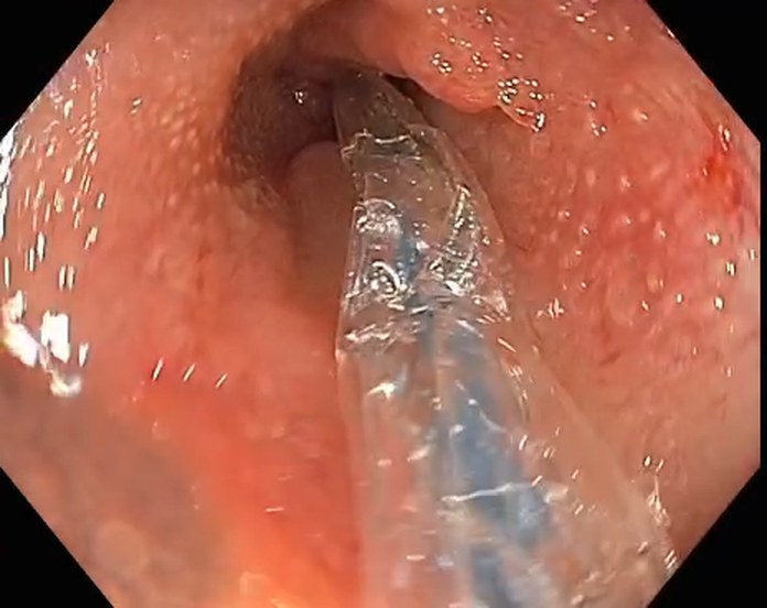
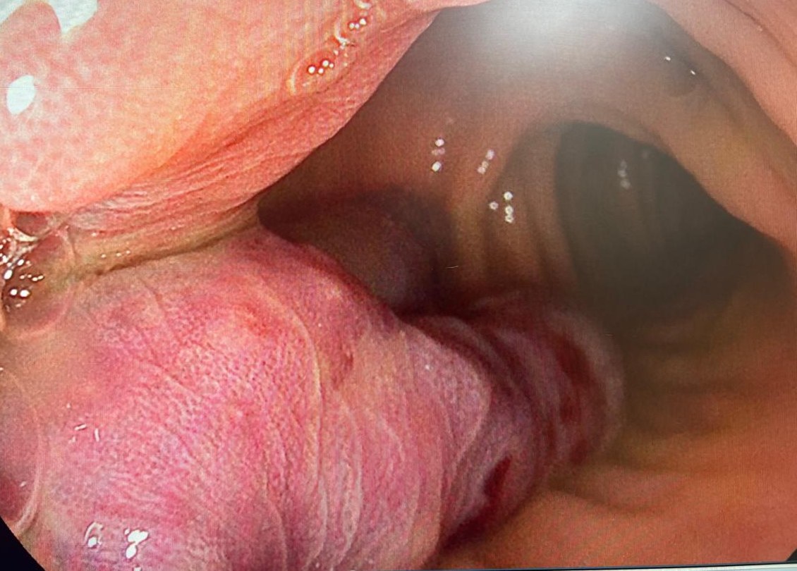
A 67-year-old woman had 30 cm of terminal ileum resected for Crohn disease in the early 1990s, then no follow-up for three decades. MR enterography in 2025 showed two 4 cm skip lesions, one at the ileocolic anastomosis and one a couple of centimeters above it. The scope could not pass the anastomosis. Endoscopy showed no inflammation. Biopsies showed mild inflammation. She started adalimumab.
Balloon dilation to 15 mm in March still did not let the scope through. She later came back with subocclusive symptoms. After a second dilation the scope still could not pass. This time the anastomotic biopsies showed low-grade dysplasia, confirmed by a second pathologist, with p53 and IMP3 positive on immunohistochemistry. She had also improved for a couple of months and gained 3 kg. The presenter did not say which treatment that followed.
The advice in the thread was surgery. One caveat: low-grade dysplasia is hard to separate from reactive change when inflammation is active. A stricture the scope cannot pass after two dilations cannot be surveyed from inside, and she did well after her first resection. Before surgery: nutrition review, albumin, prealbumin, micronutrients, and a request for a thorough pathology read of the specimen. After surgery, the options given were to continue the biologic, or to stop it and check fecal calprotectin at three months, or to reassess with colonoscopy or MR enterography at 6 to 12 months.
She agreed to resection and will stay on adalimumab. The presenter noted that this dysplasia sits inside a postoperative stricture, not a primary one. Specimen histology is due in a few weeks.
Clinical Pearl: A Crohn stricture the scope cannot pass is a stricture you cannot survey. When its biopsies show dysplasia after failed dilation, send it to surgery and ask for a careful specimen read.
3. ERCP: A Gastric Loop Made the Papilla Look Like Third-Part Duodenum
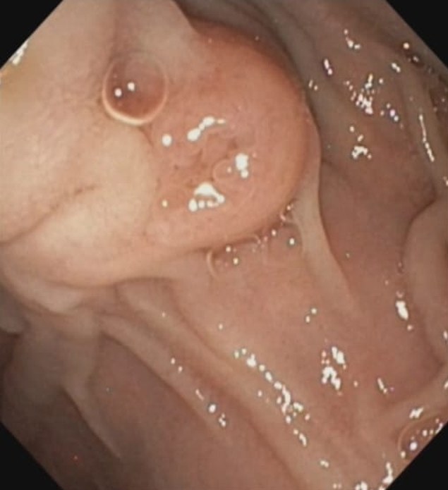
ERCP for a common bile duct stone. The papilla seemed to sit in the third part of the duodenum. The short position from the second part failed. The long position gave an en face view, but the endoscopist could not hold it and the scope kept falling back. A change from prone to left lateral did not help. Fluoroscopy showed a large loop.
The later read in the thread was that the papilla was in the second part. An earlier comment had it looking like the third part or the distal end of the second part. A loop in the stomach compressed the duodenal sweep and made the distance from the bulb to the papilla look long. In the best position reached, the scope lay tangential to the papilla. A wire would have been hard to place, and the sphincterotome would have veered to the right. Cannulation was not achieved. The endoscopist stopped the procedure to avoid injury from the scope falling back.
Suggestions for a next attempt: reduce the gastric loop first. Fluoroscopy was advised to see the loop. Right torque with the small wheel hard right, locked for stiffness if needed, is the usual first try. The same member said that, on this case, right torque may not be the reduction that works, so adjust wheel and torque by feel and by the image. Try the patient supine, and add external pressure in the epigastrium. A pediatric duodenoscope is more pliable, if one is available. A gastric overtube can keep the duodenoscope from looping in the stomach. An enteroscope was suggested by two members. One member pushed back for native anatomy, because it gives up the elevator. A published ASGE video technique for this problem was also shared.
Clinical Pearl: When the papilla seems too deep and the duodenoscope keeps falling back, suspect a gastric loop that compresses the sweep. Reduce the loop before you touch the papilla. Position comes before cannulation.
Quick Consultations
Biliary · Common duct stones, clear gallbladder, no surgery wanted
A 60-year-old woman had two small distal common duct stones. MRCP and ultrasound showed a clear gallbladder. After ERCP and sphincterotomy, she was not keen on cholecystectomy. Options for a shared decision: a generous sphincterotomy alone, sphincterotomy plus ursodiol, or a firmer push for cholecystectomy with a second surgical opinion, because the stones are presumed to form in the gallbladder. One member uses ursodiol at 10 mg/kg a day in two doses, and continues it indefinitely if the gallbladder stays. Another quoted 500 mg three times a day for 2 to 3 months, and has also used it for 12 months. The thread had no efficacy data for long-term use. One member's 84-year-old high-risk patient was refused surgery by the surgeon and the anesthesiologist, and came back three years later with cholangitis.
Liver · Anechoic cyst after hydatid surgery
An asymptomatic patient had surgery at another center for a 10 cm hydatid cyst of the liver. Follow-up ultrasound now shows a 10 cm anechoic cyst. Details of the operation were not available. One member read it as non-concerning, possibly a residual cyst if part of the hydatid was left behind. Repeat the imaging and watch for growth. No other intervention.
Stomach · Carpeted antral polyps with a normal colon and duodenum
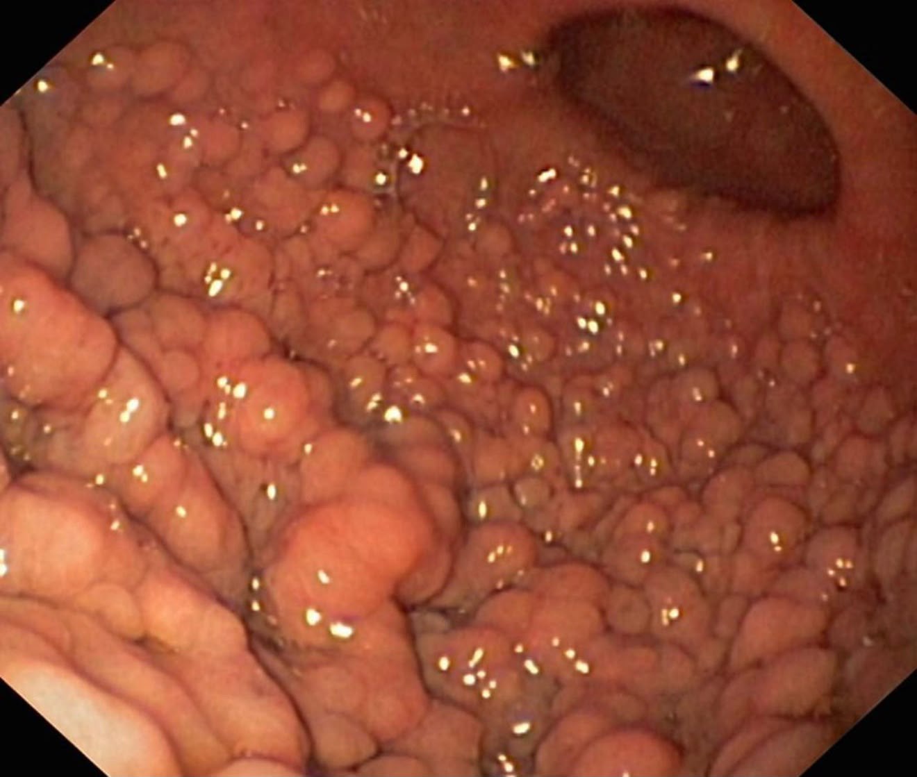
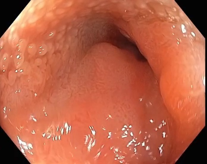
The antrum was carpeted with polyps. The proximal stomach was normal. The report was hyperplastic H. pylori gastritis, and the polyps were not adenomatous. CT still showed gastric wall thickening two months after eradication. The first question was a polyposis syndrome: colonoscopy, a careful look at the duodenum, and genetic testing if the colon is full of polyps. Colon and duodenum were clear. GAPPS was raised and set aside, because it involves the proximal stomach and these polyps were antral. Plan: rescope and rebiopsy.
Varices · Delayed bleeding from banding ulcers, INR above 2.5, platelets 35,000
One week after variceal banding, a patient came back after discharge with hematemesis. EGD showed raw post-banding ulcers. The question gave an INR above 2.5 and platelets of 35,000. One member said counts like these are common in decompensated cirrhosis and that an INR of 2.5 is not the main worry. Without active bleeding, supportive care usually works: terlipressin or octreotide, an IV PPI, fluids, and nil by mouth. For ongoing bleeding, repeat banding is an option, with a PPI, antibiotics, and terlipressin. Sclerotherapy and glue were said to work poorly here. BRTO or TIPS, as a bridge to transplant, depends on liver status and the overall condition.
What’s Next
Every case in this digest came from real conversations in the EndoCollab private WhatsApp community, 650+ practicing endoscopists discussing cases daily from every continent. Lifetime members get permanent access to the group, plus the full EndoCollab case library.