COMMUNITY DIGEST
3 featured cases and 11 quick consultations from the EndoCollab WhatsApp community between October 9 – October 23, 2026. The featured cases are a long thread on resecting flat and depressed polyps completely and setting the electrosurgical unit, a stricture of the common hepatic duct with stasis stones that members attributed to a surgical clip placed 14 years earlier, and profuse rectal bleeding that returned after a first colonoscopy. Quick consultations cover fecal elastase, resect-and-discard polls, a cardia lesion, a pancreatic head lesion, a double duct sign, pelvic floor questions, hyperplastic polyps, hepatitis C after kidney transplant, edema with a pericardial effusion in a teenager with anorexia nervosa, and gastric body biopsies.
In this issue
- Aiming for R0 on Flat and Depressed Polyps: Cold Snare, EMR, and Electrosurgical Settings
- Common Hepatic Duct Stricture with Stasis Stones, 14 Years after Cholecystectomy
- Profuse Rectal Bleeding That Returned: Hemorrhoids, a Circumferential Ulcer, and a Possible Dieulafoy
- Quick cases: Double duct sign with transient cholangitis, no EUS on site; Hyperplastic polyps and the surveillance interval; Resect and discard for polyps under 10 mm: two informal polls; Hepatitis C reactivation in a fresh kidney transplant; Edema and a pericardial effusion in a teenager with anorexia nervosa; Low fecal elastase: valproate, false positives, and gastric bypass; Pancreatic head lesion on EUS: mass or groove pancreatitis?; Dyssynergic defecation with a rectocele: proper biofeedback before surgery; How one fellowship taught the digital rectal exam for pelvic floor dysfunction; Cardia lesion after dyspepsia: retch gastropathy or something else?; Gastric body with hard, slippery biopsies: gastritis, or more?
Featured Cases
1. Aiming for R0 on Flat and Depressed Polyps: Cold Snare, EMR, and Electrosurgical Settings
A member asked when to restart anticoagulation after removing polyps of 1 cm or less, citing a talk on cold snare polypectomy in anticoagulated patients that departs from BSG guidance. One member answered: for polyps under 1 cm cut with no heat, within 24 hours (some say the night of the procedure); for polyps cut with heat, 48 to 72 hours. The member assumed a DOAC, and said that if you can cut without electrocautery, pit pattern and morphology do not matter. A second member agreed. No guideline was cited.
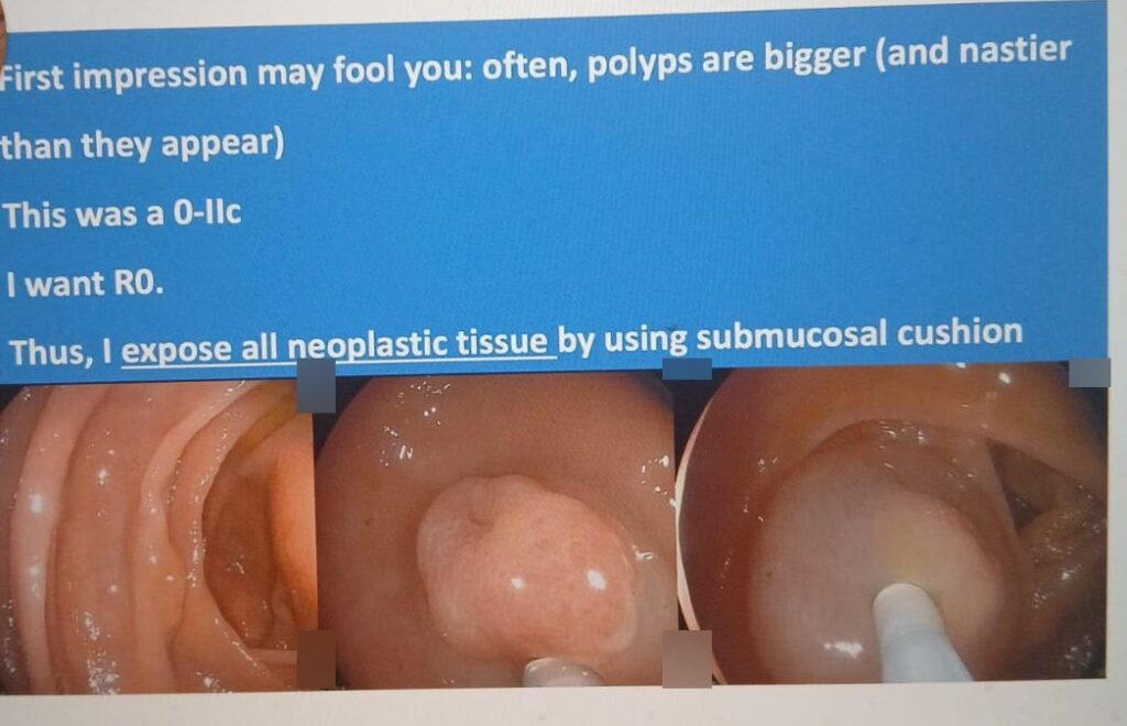
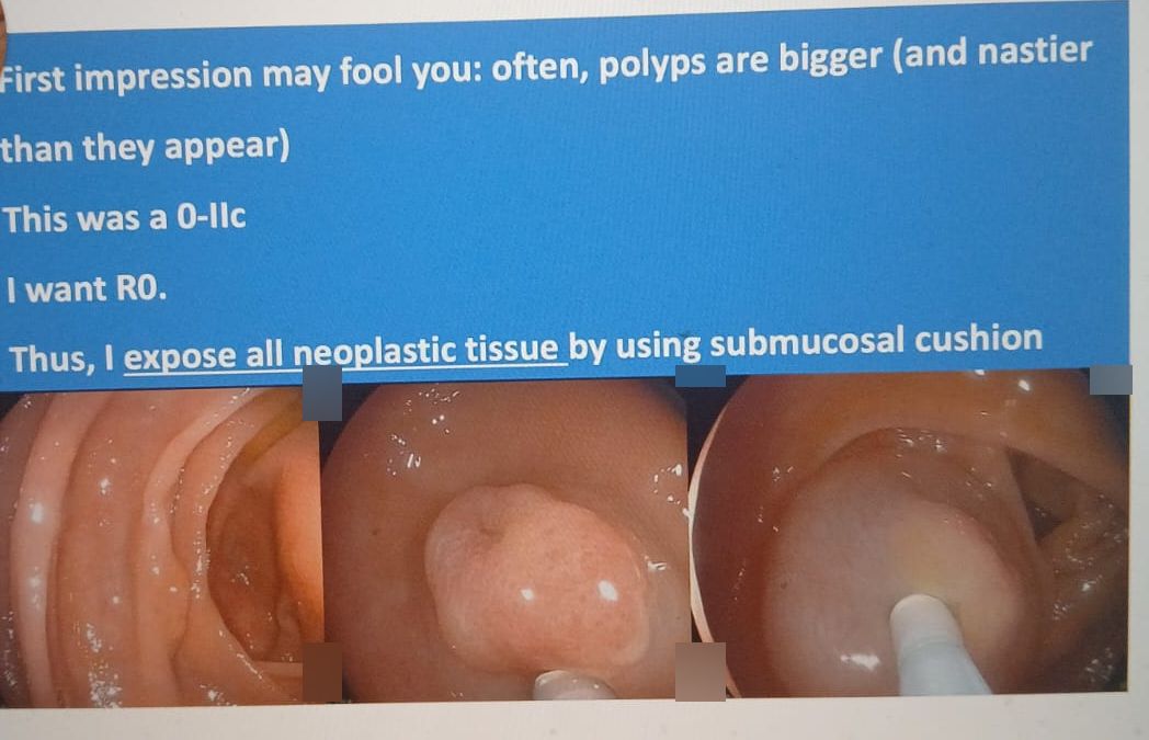
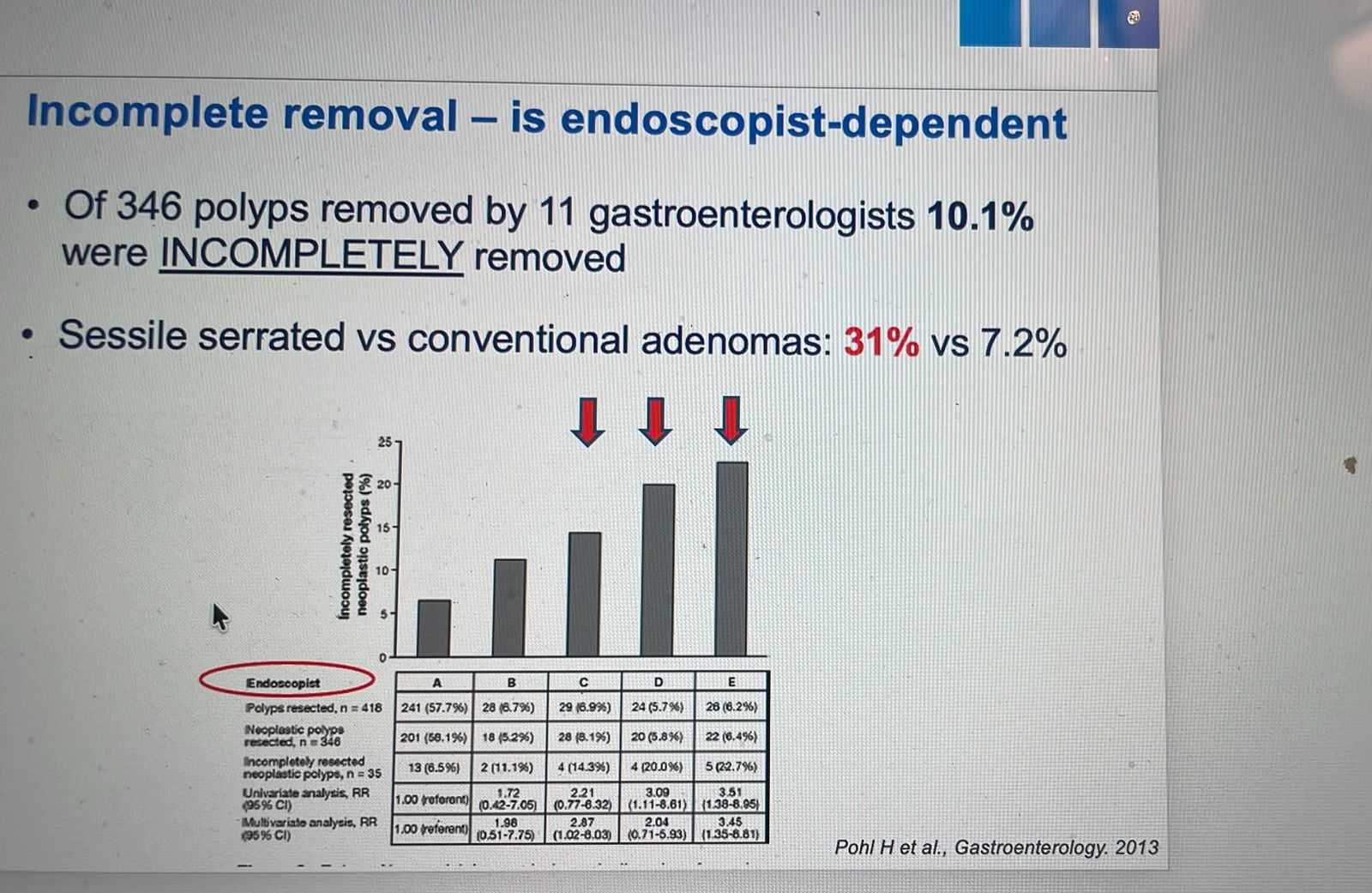
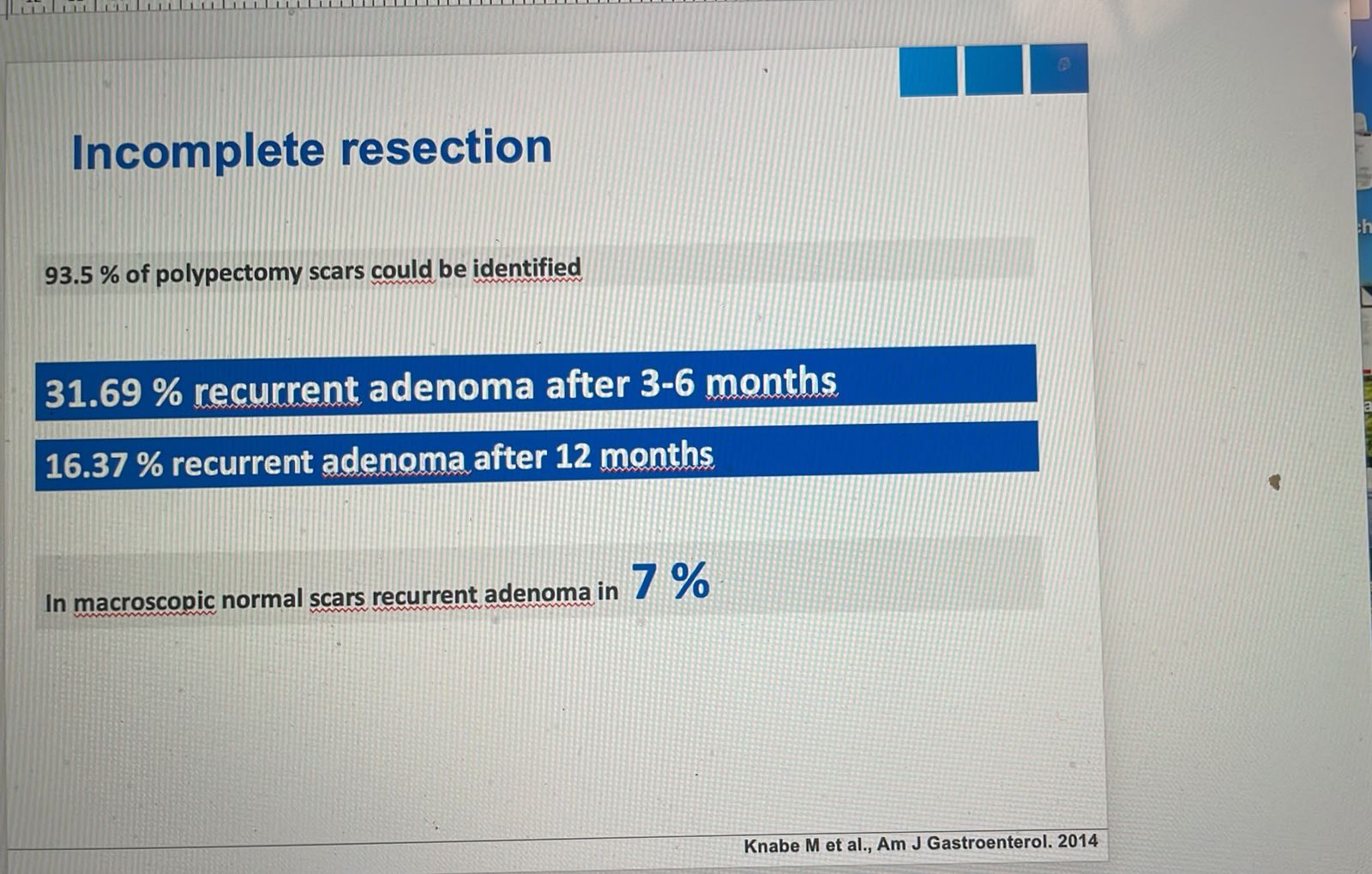
A follow-up asked whether a 1 cm polyp with a IIc component can still be taken with a cold snare, and what ERBE settings suit a circumferential incision on a flat lesion. Prof. Klaus Mönkemüller answered that the aim is complete removal of neoplasia, R0. A IIc lesion has a higher chance of advanced neoplasia, and he knows of too many cases and studies where plain polypectomy, cold or hot, left flat or depressed lesions incomplete. He put recurrence after plain polypectomy at about 10 to 15% and called that bad. His slides: 10% of polyps incompletely resected, more often serrated lesions (31% vs 7%), and 32% recurrence in a study of larger lesions. His advice: use the technique that makes a flat polyp more visible, defines its borders, and cuts wide enough laterally and deep enough vertically. Submucosal cushion with EMR, or underwater EMR, have the highest yields of R0.
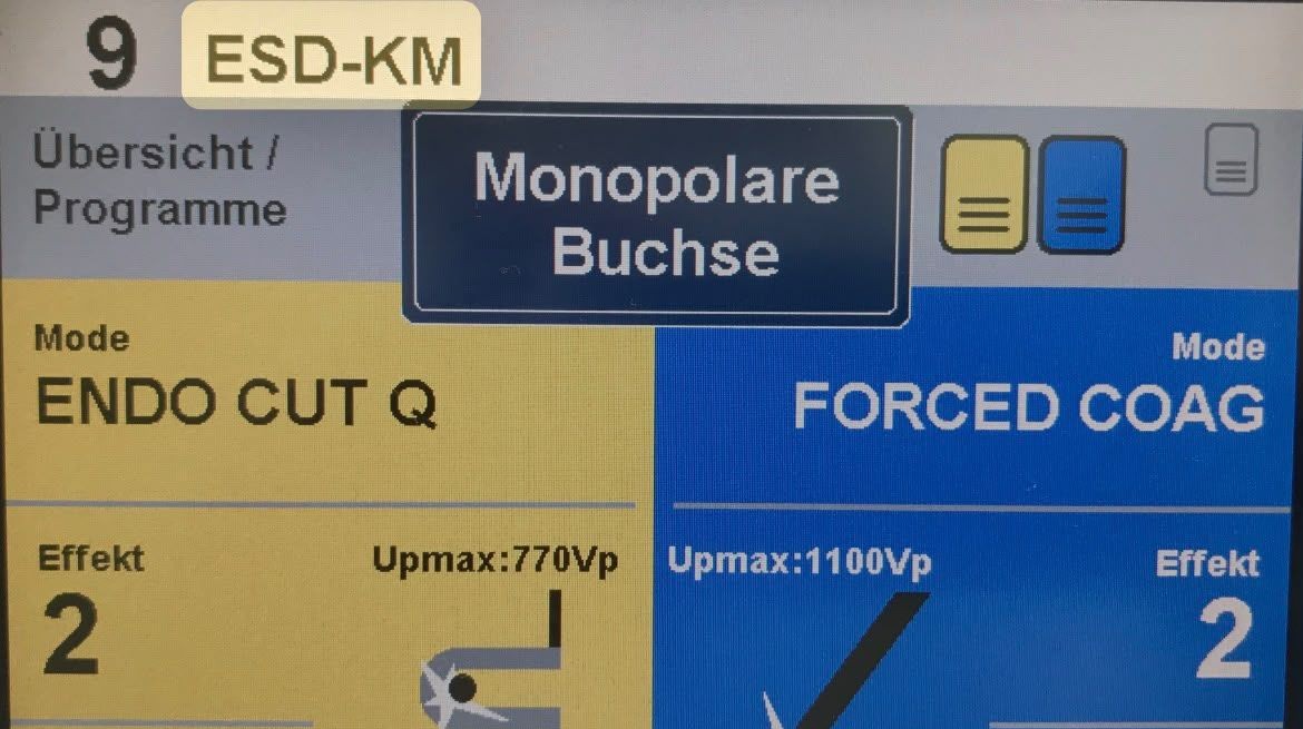
For the yellow pedal he recommended his unit’s settings, which a member read as 2-3-3, and said they work for EMR too. A member who uses 2-1-6 or 3-1-6 was told his settings were perfect for mucosectomies, because the longer interval (6) suits them better. ERBE combines effect (strength of the current), duration (length of each pulse) and interval (the cooling pause). Without ERBE’s microprocessor, effect is the key setting (1 pure cut, 3 a mix of cut and coagulation, 6 more coagulation). He likes effect 2, and controls duration with the length of pedal taps and interval with the pauses off the pedal.
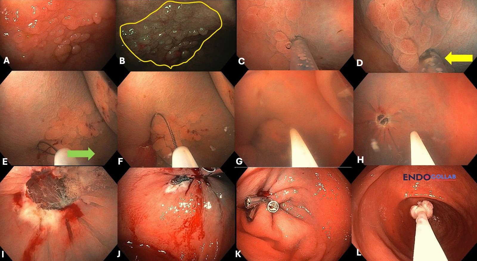
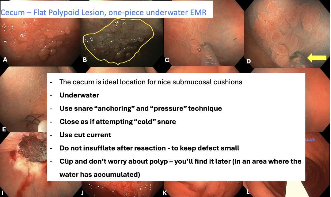
On October 21, a member watching the EndoCollab lecture on the size limit of EMR asked why a short hot cut, a cold closure, then another hot cut would lower perforation risk. Another member said it depends on the settings: uninterrupted pure cut or coagulation spreads heat, so tap and rest, but settings with built-in pauses remove the need to lift your foot. Prof. Mönkemüller added that ERBE cannot replace your judgement of the visible cutting effect, especially in the cecum, where a poor cushion on a 20 mm flat polyp and constant pressure on the pedal may end in perforation. When the cushion is not ideal, start with a controlled initial cut and watch how the tissue responds. He posted another flat cecal lesion, 20 mm, that came out in one piece with cushion plus underwater technique and left a tiny wound. With cushion and water, he said, the unit’s settings lose their meaning, because the water dissipates the current. A new lecture on complex colon polyps is in EndoCollab, and a tips-and-tricks lecture on electrocoagulation should be ready by December.
Clinical Pearl: The aim is R0, not just removal. A IIc lesion is more likely to hide advanced neoplasia, and plain cold or hot polypectomy leaves some flat or depressed lesions incomplete. Prof. Mönkemüller said a submucosal cushion with EMR, or underwater EMR, gives the highest R0 yields. Treat the unit's settings as a starting point: watch the tissue respond; Prof. Mönkemüller advises tapping the pedal rather than holding it, though one member noted that settings with built-in pauses make this less critical.
2. Common Hepatic Duct Stricture with Stasis Stones, 14 Years after Cholecystectomy
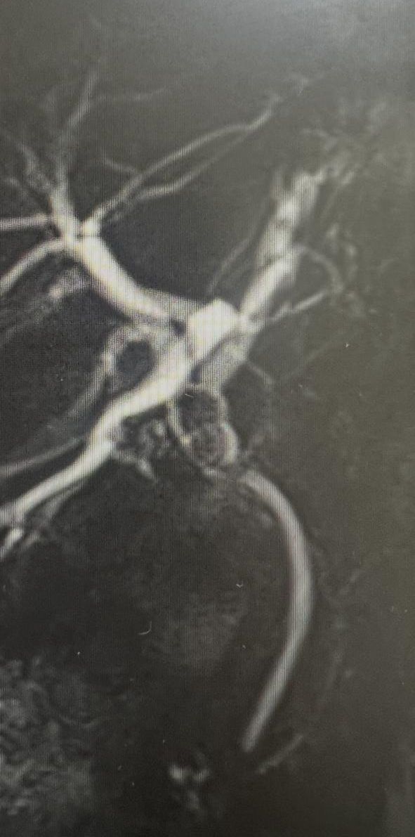
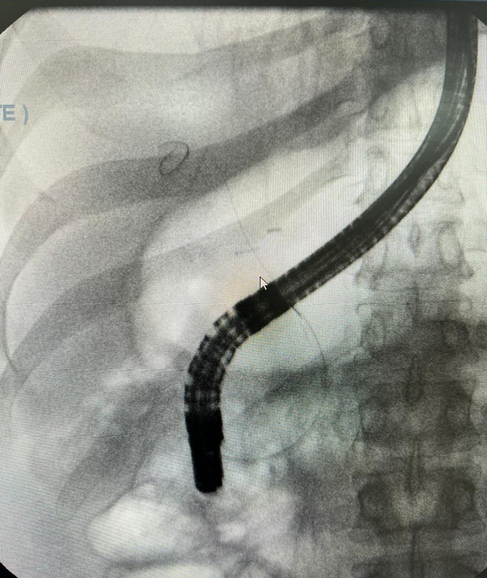
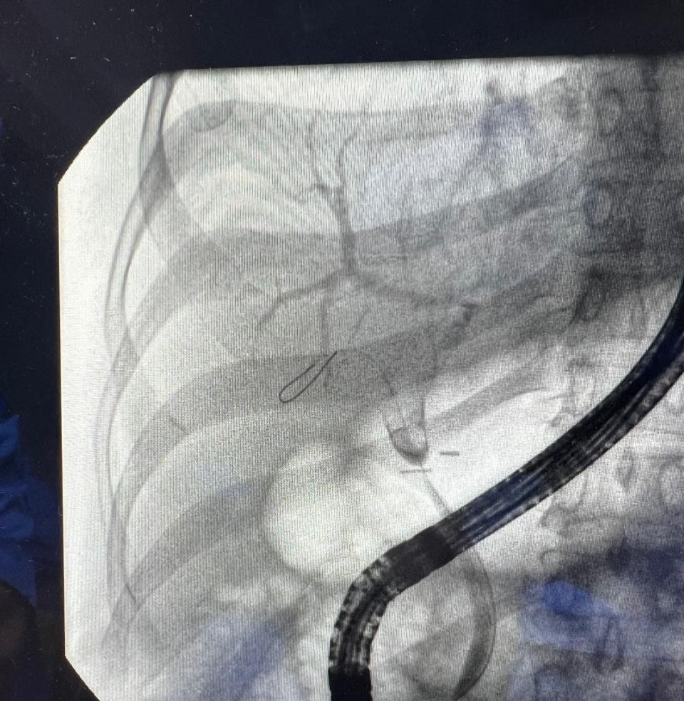
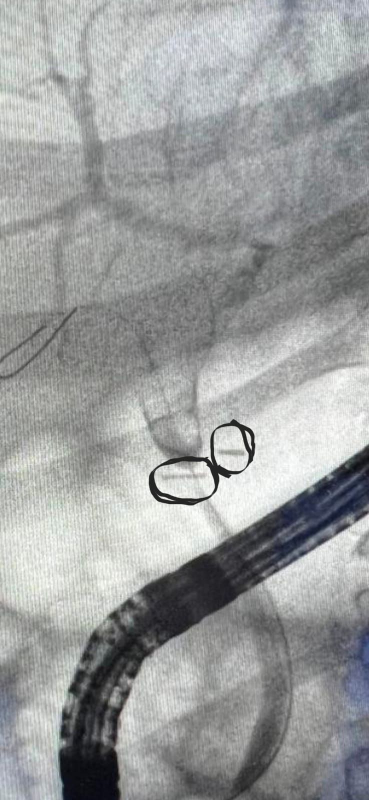
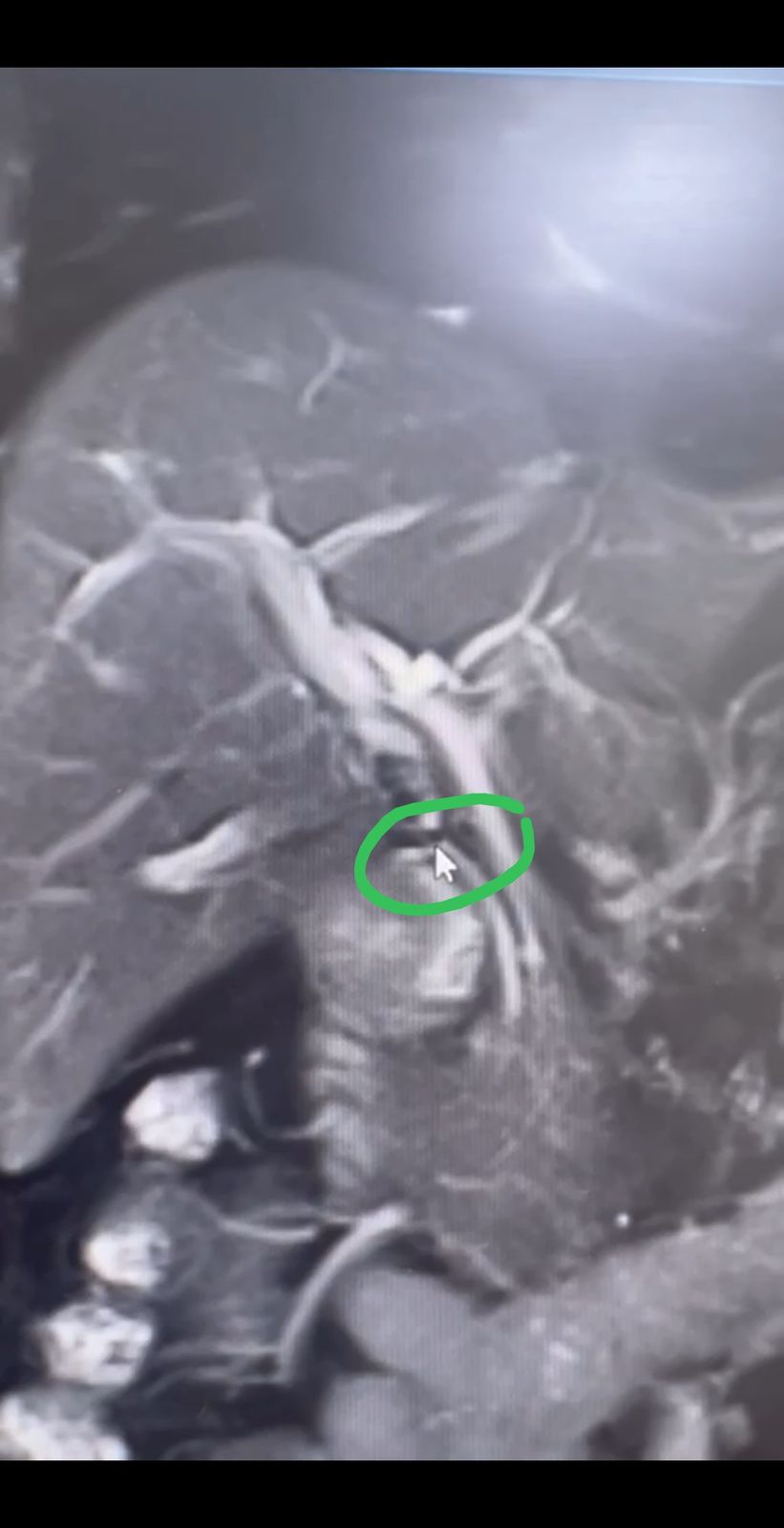
A 40-year-old man with chronic hepatitis B (HBeAg negative, HBV DNA 925 IU/ml) came with a week of itching and no abdominal pain. ALT was 272, AST 113, ALP 471, bilirubin normal, ultrasound normal. MRCP showed a stricture of the common hepatic duct with two gallstones. At ERCP the guidewire crossed the stricture easily, but a 7 French x 10 cm Amsterdam straight stent would not pass. He had a laparoscopic cholecystectomy 14 years ago. Liver tests were normal in the 2024 and 2025 records, and an ultrasound in 2024; in February and March AST and ALT rose from about 50 to 150. He took tenofovir alafenamide for two months and stopped.
One member called the stricture very focal, advised cholangioscopy to be sure, and thought it likely inflammatory given how short and focal it was. He read the stones as stasis stones from the stricture, and the fluctuating enzymes as stones blocking and unblocking. He then circled two surgical clips on the fluoroscopy still, one over the problem area, found the same clip on the MRI, and said: “I think that’s your diagnosis.” The presenter, looking back, saw the clip clearly and thought it had partly caught the duct, but was puzzled why it took 14 years. The reply: the clip did do something, with dilatation, stasis, sludge and stones, and was probably fired across the duct unnoticed, tight enough for stasis but not for complete blockage. Secondary biliary cirrhosis is a risk. Another member said cholangioscopy would be for academic interest only, since the cause was clear.
On treatment, one member would dilate with a balloon, starting at 4 mm and going 4, 6, 8 (Soehendra dilators were an alternative, but they top out at about 3.8 mm, similar to a 4 mm balloon). The stones must come out, by electrohydraulic lithotripsy through a cholangioscope or by dilating enough for them to pass. His preferred option was an 8 mm fully covered metal stent for 3 to 4 weeks: the clip would hold it, radial force would act for weeks rather than minutes, and it may break up the stones. Leave the distal 1 cm outside the papilla so it is easy to grasp later. He likened the stricture to a post-transplant anastomotic stricture (about 85% success at 12 months, in his expectation), said these can recur, follows patients yearly with liver tests and MRCP, and discharges at 3 years without recurrence. Surgery means hepaticojejunostomy, which he would avoid in a 40-year-old unless the endoscopic route fails.
The presenter reasoned that since the 7 French stent would not cross, the opening is under 2.33 mm, though the wire passed smoothly. He planned a 4 mm balloon, 2 cm long, with a 4 French tip, and a member replied that a deflated balloon should cross. Whether it was dilated, and how the patient did, was not reported.
Clinical Pearl: When MRCP shows a short, focal stricture of the common hepatic duct with stones after a cholecystectomy, ask whether a surgical clip is crossing the duct. In this thread the clip was missed at first and then identified on both fluoroscopy and MRI. The stones were read as stasis stones. Balloon dilation or a fully covered metal stent were the endoscopic options discussed, with hepaticojejunostomy as the fallback. The outcome was not reported.
3. Profuse Rectal Bleeding That Returned: Hemorrhoids, a Circumferential Ulcer, and a Possible Dieulafoy
A member shared a colonoscopy video showing hemorrhoids plus a circumferential rectal ulcer in a 70-year-old woman with profuse lower GI bleeding and no history of diabetes or heart disease. Her hemoglobin had dropped from 9 to 4 g/dl in 24 hours. The first reply asked about rectal prolapse and about a pressure ulcer from fecal impaction. The presenter said hard impacted stool was everywhere at colonoscopy, so that first impression fit, and asked whether it could be anything else. No further answers came that day.
Eight days later the patient returned with profuse bleeding and collapsed. An urgent sigmoidoscopy video was posted, and the presenter’s impression was a rectal Dieulafoy lesion. One member read it as a rectal ulcer and hemorrhoids, then saw a pulsatile lesion that seemed to arise from the earlier area of ulceration. He would not call it a Dieulafoy as such, but it looked like the culprit for the bleed. He assumed a full colonoscopy had shown nothing suspicious beyond the rectum, and said that either way the lesion should be injected and clipped. Another member said it looked like a rectal Dieulafoy. The presenter confirmed that nothing was found beyond the rectum at the earlier colonoscopy and that CT mesenteric angiography was normal. The thread ended there, with no report of treatment or outcome.
Clinical Pearl: In recurrent massive rectal bleeding, hemorrhoids and a rectal ulcer may not be the whole story. Members split between “ulcer and hemorrhoids” and “rectal Dieulafoy” for the lesion seen at the second look, and the only treatment suggested was to inject and clip it. The colon beyond the rectum was clear and mesenteric CT angiography was normal. The outcome was not reported.
Quick Consultations
Biliary · Double duct sign with transient cholangitis, no EUS on site
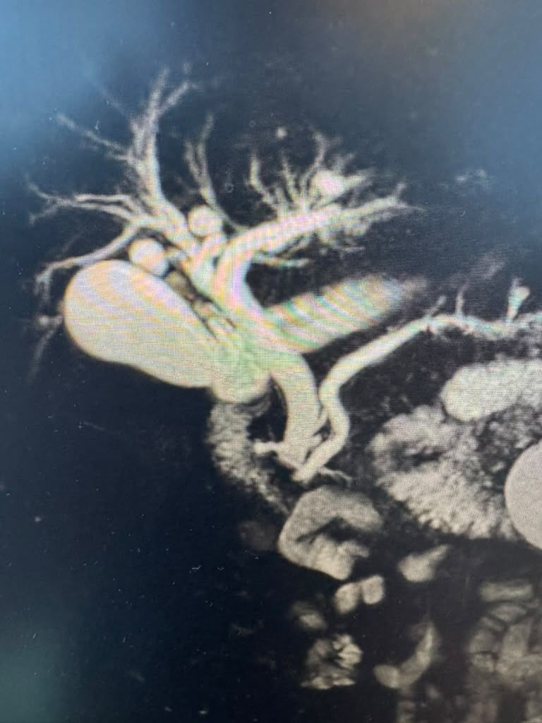
At a center without EUS, a patient with a double duct sign had transient cholangitis symptoms and improved on antibiotics. Bilirubin was normal. MRCP showed enhancement at the distal common bile duct, read as a possible ampullary mass or fibrosis. Transfer for EUS, or ERCP now? One member said an MRCP showing an ampullary mass is a clear indication for ERCP: look directly, biopsy, decompress and drain, then add EUS for the extent. From the image he could not say a mass was definite. He saw an abrupt cut-off in the distal duct, listed distal cholangiocarcinoma among the possibilities, thought a pancreatic head lesion unlikely to obstruct the distal-most duct so neatly, and kept an ampullary lesion on the list. With an INR of 2.2, he suggested vitamin K and FFP and a stat INR just before the procedure, sampling if it corrects. Otherwise, wire and stent with no sphincterotomy and no biopsies. Outcome not reported.
Colon · Hyperplastic polyps and the surveillance interval
A member asked whether hyperplastic polyps in the sigmoid count toward the number used for the surveillance interval, for example more than five hyperplastic polyps and a repeat in three years. One member said no: unless there are more than 20, hyperplastic polyps can be disregarded. With more than 20, think of a hyperplastic and serrated polyposis syndrome, which is not necessarily genetic, treat the patient as high risk and screen yearly. The one thing to check is that the pathologist is not calling a sessile serrated polyp hyperplastic. The clues are size, right-sided location, and an endoscopic look that does not suggest a hyperplastic polyp. He also believed, from memory, that USPSTF calls for a 5-year repeat for left-sided hyperplastic polyps over 10 mm and a 3-year repeat for a sessile serrated polyp over 1 cm proximal to the sigmoid.
Colon · Resect and discard for polyps under 10 mm: two informal polls
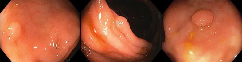
Grat ran two informal polls. On resect-and-discard for colon polyps under 10 mm, 3 members voted yes and 62 no. On sending all polyps under 10 mm for histopathology, 66 voted yes and 3 no (counts at the time of the export). In the discussion, one member always uses virtual chromoendoscopy to decide, and thought a serrated or adenomatous polyp under 10 mm with no suspicious pattern, judged with high confidence, can be resected and discarded. Another resects only what looks adenomatous or uncertain and leaves non-concerning, hyperplastic-looking polyps in place. He believes resect-and-discard is accepted in the UK, applies it only outside polyposis syndromes, and adopted his current approach four or five years ago. A third does not resect hyperplastic-looking diminutive rectal polyps.
Hepatology · Hepatitis C reactivation in a fresh kidney transplant
A fresh renal transplant, admitted with sepsis, likely had hepatitis C reactivation after remote treatment, probably with interferon. Treat now or wait? One member said this is not the time: treating hepatitis C is elective, and it should wait until the patient is out of the woods. When the time comes, the options depend on renal function and cirrhosis. His preferred regimen is Epclusa if eGFR is above 30 ml/min (pangenotypic, success above 97%). With marked renal impairment, Mavyret. If the genotype is not 2 or 3, Harvoni. He noted that Epclusa and Harvoni interact least with tacrolimus, cyclosporin and mTOR inhibitors. A second member agreed fully. The presenter had been considering Epclusa, and renal function was good.
Nutrition · Edema and a pericardial effusion in a teenager with anorexia nervosa
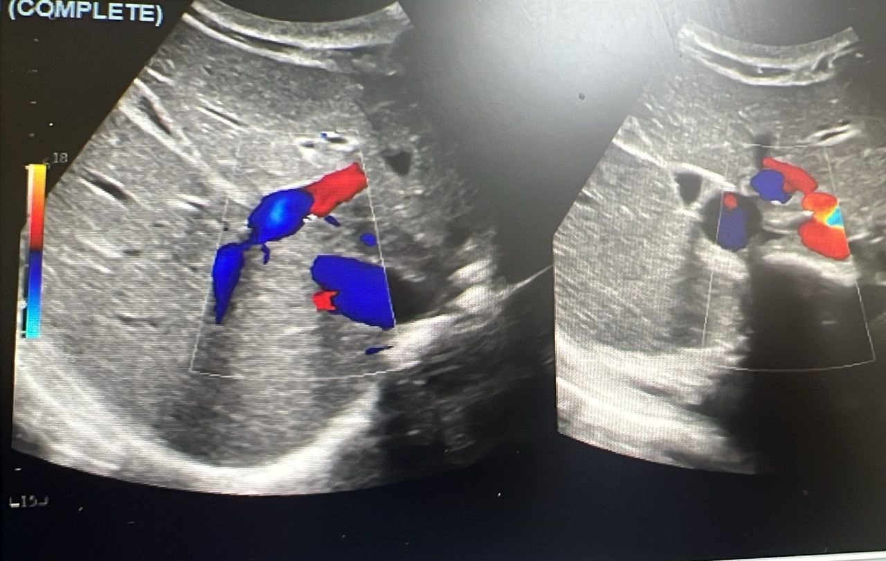
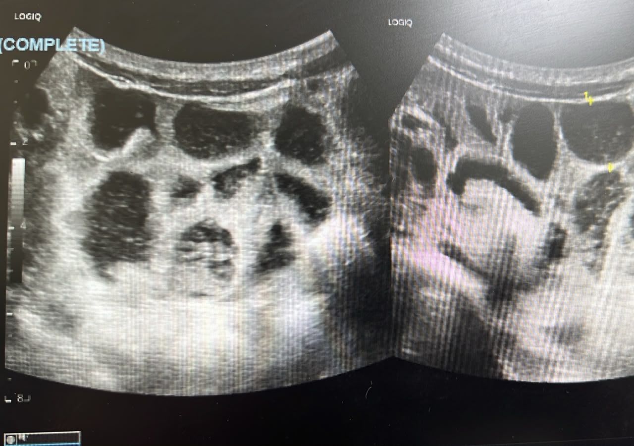
A 15-year-old girl diagnosed with anorexia nervosa elsewhere had lost weight from 67 to 33.5 kg in a year and a half and was on tube feeding. She came with pedal edema and abdominal distension. Albumin had risen from 32 to 35.3. Ultrasound showed edematous small bowel loops and free pelvic fluid, urine showed no protein loss, and echo showed a non-collapsing pericardial effusion of 5 to 10 mm with a normal ejection fraction. One member found the picture hard to explain with an albumin that good, listed connective tissue disease, sarcoidosis and TB among the possibilities, and advised celiac testing, gastroscopy and colonoscopy (MR enterography if she refuses), with the cardiologist to investigate the effusion. The presenter reported a chest X-ray without TB stigmata, hemoglobin 14.5, platelets 245 and normal electrolytes. Prof. Mönkemüller added Addison disease to the differential. Another member said refeeding syndrome would top the list if the features began after feeding started, advised leaving the effusion alone, and suggested safety-netting the family. The edema was decreasing, with a repeat ultrasound planned in 3 weeks and an echo in 4. The diagnosis stayed unresolved; if she improves, the presenter will treat it as malnutrition-related fluid overload.
Pancreas · Low fecal elastase: valproate, false positives, and gastric bypass
A member asked whether valproate could cause pancreatic insufficiency in a patient with chronic diarrhea and low fecal elastase on two occasions three years apart. No direct answer on valproate was given in the thread; one member asked about fecal fat. Another said fecal elastase has many false positives: the pancreas secretes little at some times, such as during fasting, so the result can be very low and lead to enzymes the patient does not need. Clinical context and nutritional assessment come first, and secretion is not function (he named the 13C mixed triglyceride breath test for function). A second member said low elastase is common after gastric bypass and asked when to treat. The first member explained that vagotomy, antrectomy and duodenal exclusion remove stimulation of the pancreas, and that in a Roux-en-Y the few enzymes secreted meet the food out of step. He advised checking nutrition (weight, albumin, micronutrients, fat-soluble vitamins) and taking enzymes at the start, during and at the end of the meal. His suggested Creon 25,000 schedule: 2 capsules at breakfast, 1 mid-morning, 3 at lunch, 1 in the afternoon, 2 at dinner. This thread began on October 8 and was not covered in Issue 12.
Pancreas · Pancreatic head lesion on EUS: mass or groove pancreatitis?
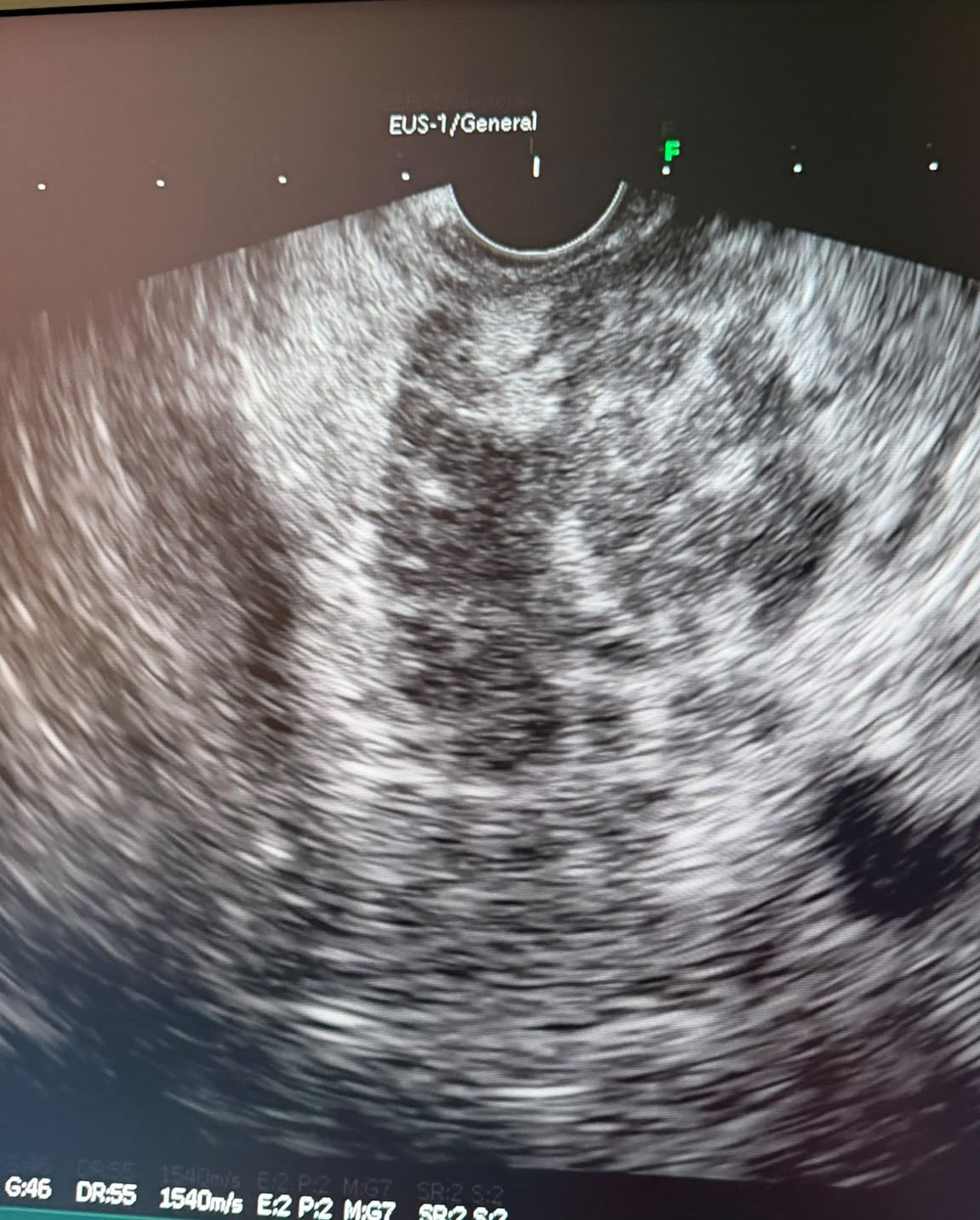
A patient referred for incidental choledochal dilation, with no complaints and no history of pancreatitis, had a 33 x 39 mm lesion in the pancreatic head on EUS. The presenter’s differentials were SCN and groove pancreatitis. One member asked for more than one MRI sequence. From the image alone he thought chronic or resolving pancreatitis, depending on history, but found it more concerning for a real mass, although groove pancreatitis is possible. The presenter had only an MRCP and will arrange a triphasic MRI. The advice: do the proper contrast MRI, and if doubt remains, repeat the EUS in 6 weeks. Another member agreed. Outcome not reported.
Pelvic floor · Dyssynergic defecation with a rectocele: proper biofeedback before surgery
A 41-year-old woman had difficulty evacuating and a normal colonoscopy, with anorectal manometry, endoanal ultrasound and MR defecography done. A colorectal surgeon suggested rectocele repair and possibly repair of an anterior external sphincter defect. She has no fecal incontinence, splints the perineum and feels a bulge. The presenter doubted that repair makes sense if dyssynergic defecation is not corrected. She had 10 sessions of biofeedback at a center used to treating urinary incontinence, with perineal electrodes and later a vaginal probe. Two members said that is not proper anorectal biofeedback. One advised manometry-based biofeedback, with balloon insufflation for better perception, and thought surgery would not improve defecation since the rectocele is small. He relayed an expert protocol of 6 sessions 1 to 2 weeks apart, then reinforcement at 3 months, 6 months and 1 year. Another member said sphincter repair is not needed without symptoms, that rectocele repair may make her worse, and that Botox to the puborectalis is not first line: the evidence is anecdotal and he recalled about 40% sustained benefit at 12 months. The presenter chose to try proper biofeedback first.
Pelvic floor · How one fellowship taught the digital rectal exam for pelvic floor dysfunction
Asked for a learning resource on the digital rectal exam for pelvic floor dysfunction, a member described what a visiting fellowship in a neurogastroenterology department taught: explain everything to the patient, start by feeling anal tone, sweep the rectum for megarectum, then, with a hand on the abdomen, ask the patient to bear down, watching whether the abdominal muscles contract appropriately and feeling for a rectocele, and finally ask the patient to squeeze your finger.
Stomach · Cardia lesion after dyspepsia: retch gastropathy or something else?
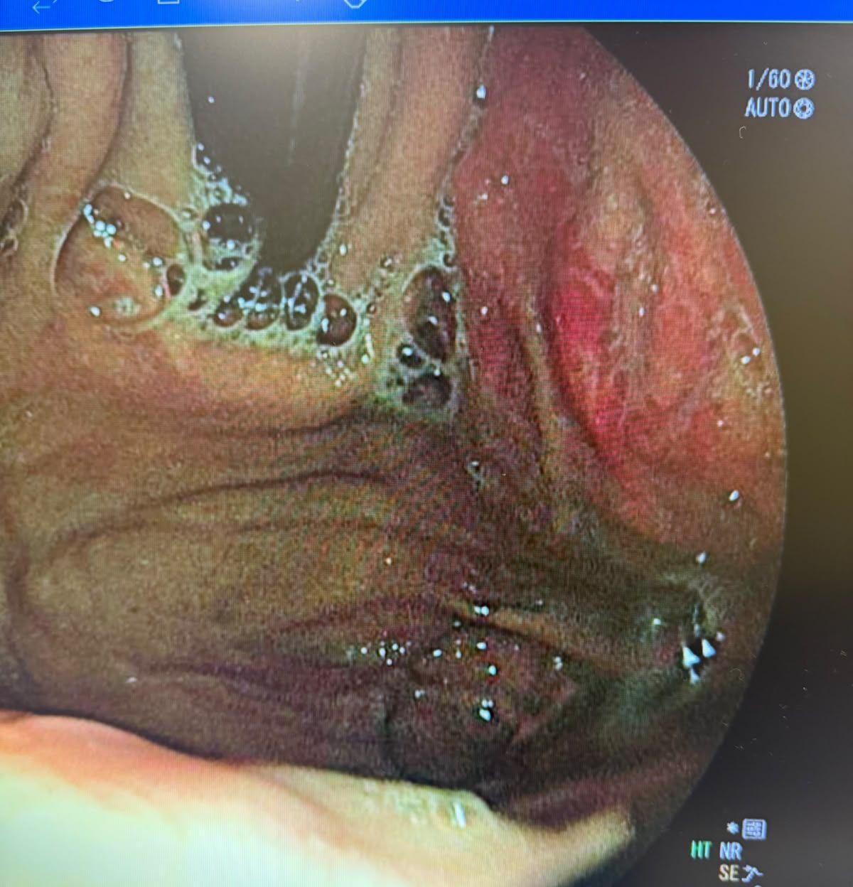
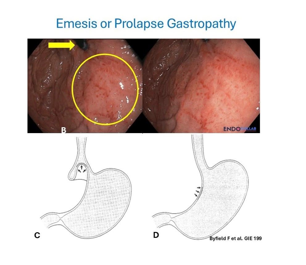
A 46-year-old woman with dyspepsia and no B symptoms had a cardia lesion whose biopsies were negative for malignancy. A control EGD was planned, but the presenter wanted to know whether it is really benign. One member said such a lesion is not unusual after persistent nausea and vomiting: the cardia is engorged with petechiae and biopsies are non-revealing. If the history fits, repeat in 4 weeks and the area should look unremarkable. Others said prolapse gastropathy or retching. Prof. Mönkemüller agreed it could be emesis or prolapse gastropathy and pointed to a newsletter article on the topic. Another member also sees this pattern in H. pylori gastritis but found traumatic fundal gastropathy more typical here. The presenter then reported that the biopsy also showed H. pylori. One member suggested a Cameron lesion, with a remote possibility of malignancy.
Stomach · Gastric body with hard, slippery biopsies: gastritis, or more?
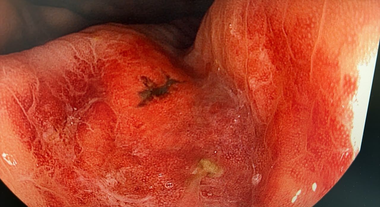
A 60-year-old with left-sided abdominal pain had an OGD in which biopsies felt slippery and hard, especially between folds. The lesion was in the mid body; the presenter thought the pit pattern looked adenomatous (Kudo IIIs) and noted an ulcer with pigmentation. One member said prolapsing gastropathy, with no need for biopsy, and asked for an NBI image; another said gastritis with erosive changes and, after the extra images, kept that answer. Another member said that if the photo is the gastric body, the glandular pattern is the normal fundic gland, or honeycomb, pattern, because the glands are vertical, unlike the oblique antral glands. What is not visible is the collecting venule pattern, which could point to H. pylori. The presenter found the answers reassuring.
What’s Next
Every case in this digest came from real conversations in the EndoCollab private WhatsApp community, 650+ practicing endoscopists discussing cases daily from every continent. Lifetime members get permanent access to the group, plus the full EndoCollab case library.