COMMUNITY DIGEST
3 featured cases and 8 quick consultations from the EndoCollab WhatsApp community between October 24 to November 3, 2026. A post-cholecystectomy patient with a collection by the cystic duct stump and an MRCP that showed no stones, new duodenal nodules a decade after an EMR, and a rescue plan for a hepaticogastrostomy stent that stopped draining. Plus a follow-up to the 14-year-old clip from Issue 13.
Since the last digest: 221 messages from 30 members, and 13 cases posted for a second opinion.
In this issue
- Liver Tests at 700, Three Weeks after Cholecystectomy: Wait, EUS, or ERCP?
- New Duodenal Nodules, 10 Years after a Duodenal EMR
- A Migrated Hepaticogastrostomy Stent with No Wire Access
- Follow-ups: Why a cholecystectomy clip can cause trouble 14 years later
- Quick cases: Gastric bypass for GERD and gastroparesis in one operation?; Refractory shock at night with no IR or surgical backup; A sigmoid ulcer after diarrhea that started before a trip; A 1.5 cm liver cyst, hepatomegaly, and a family history of cholangiocarcinoma; Probable main-duct IPMN in an 84-year-old: EUS if surgery is off the table?; Rectal bleeding with prolapse and a probable solitary rectal ulcer; Pseudo-obstruction and pneumatosis in myeloma, with amyloid on biopsy; Aphthous ulcers from the esophagus to the bulb, calprotectin 450
- Community notes: Three questions one senior member asks himself before advisi…; One member's experience with five marginal ulcers at the gas…
Featured Cases
1. Liver Tests at 700, Three Weeks after Cholecystectomy: Wait, EUS, or ERCP?
First reply in 1 minute. 4 colleagues weighed in.
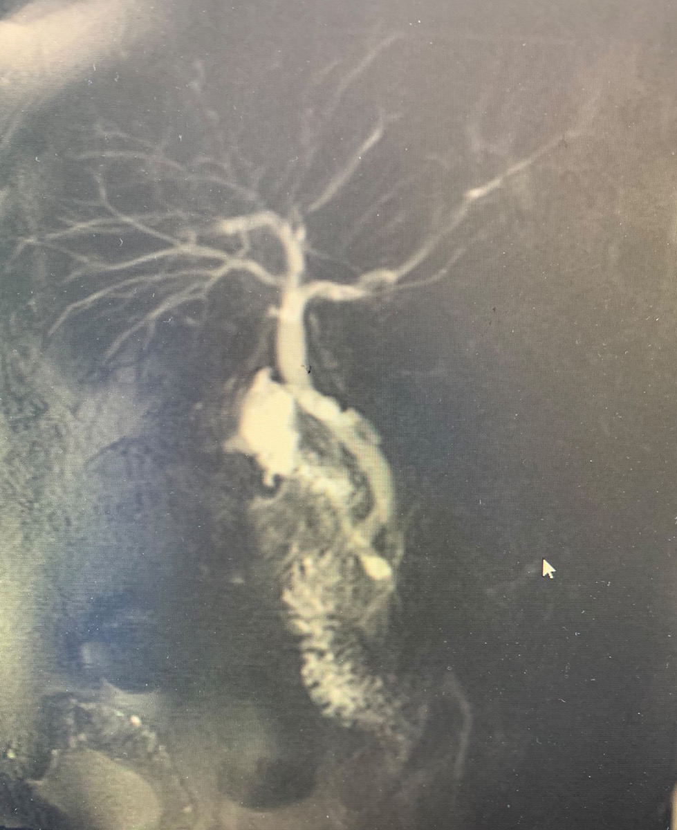
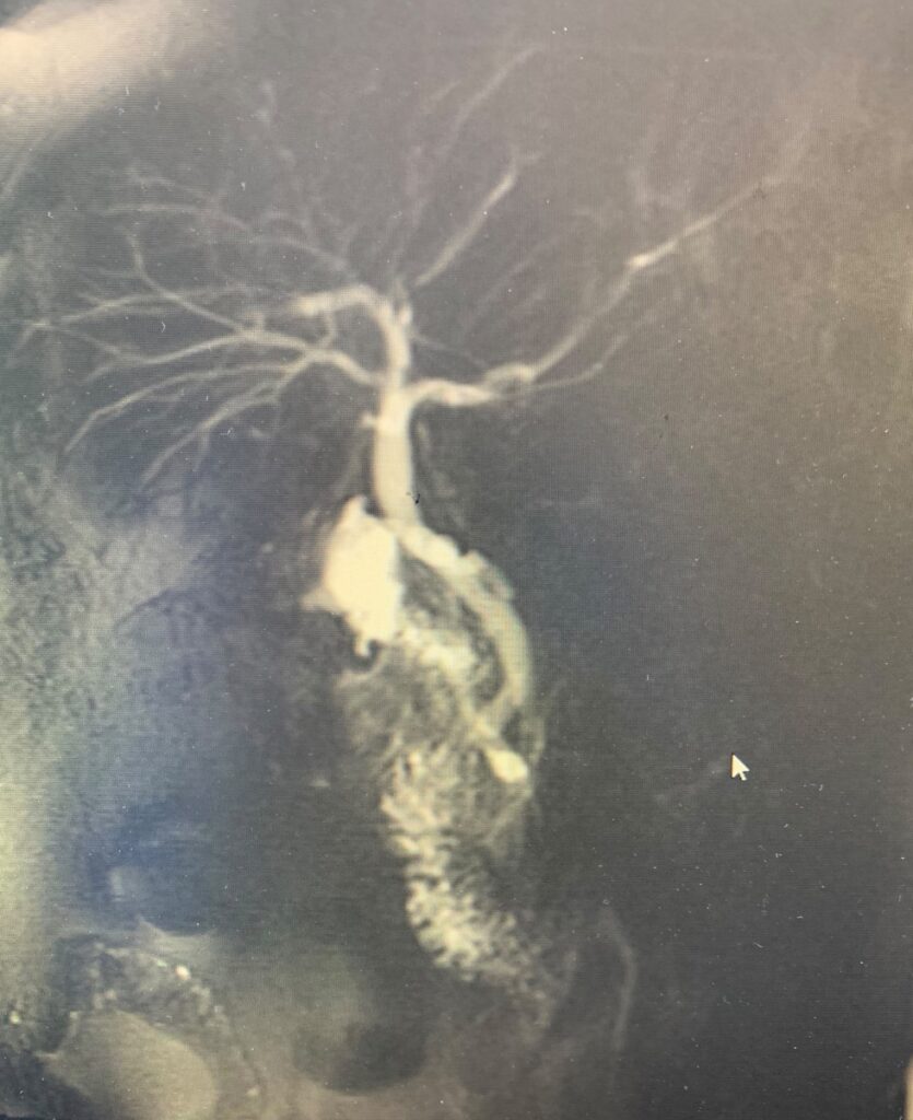
Three weeks after a laparoscopic cholecystectomy, with 5 mm stones in the gallbladder on pathology, a patient came to the emergency department with right upper quadrant pain. Bilirubin was 4.3, AST and ALT were around 700, and alkaline phosphatase was 374. There was no fever and the white count was normal. CT showed no biloma. MRCP showed a 6 mm common bile duct with no stones, and a cystic duct stump or gallbladder remnant. The radiologist's read included a small hematoma. EUS was not available.
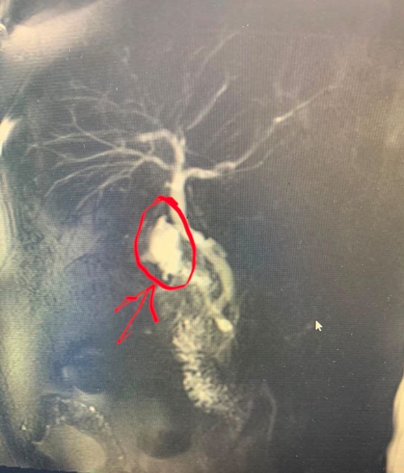
The question was whether ERCP was indicated. The first reply came a minute later, asking about the bright area on the 3D reconstruction, circled in the second image. It looked like a collection. That member read it as a leaking stump, possibly after a subtotal cholecystectomy, and favored ERCP with sphincterotomy, with a surgeon to assess the remnant.
A second member pointed out that small distal stones can be missed on MRCP soon after cholecystectomy, and said his center would go to ERCP. A third took a more measured line. If the patient was pain free and the collection was a hematoma, repeat the liver tests the next day. If they improved, assume a stone had passed. If they did not, get an EUS. If it was a biloma, do ERCP with sphincterotomy, with a stent if needed.
The presenter went ahead with ERCP. The night before, the patient developed cholangitis, with a lipase of 2,000. ERCP cleared sludge, a tiny stone, and pus from the duct. There was no leak. The patient was symptom free that evening, when the presenter posted the outcome and thanked the group.
Clinical Pearl: Soon after cholecystectomy, a normal-caliber duct on MRCP does not rule out a small distal stone. In a symptomatic patient with a collection by the stump, ERCP with sphincterotomy treats both a retained stone and a stump leak.
This case was posted and answered in our private WhatsApp group of 650+ endoscopists.
2. New Duodenal Nodules, 10 Years after a Duodenal EMR
First reply in 21 minutes. 5 colleagues weighed in.
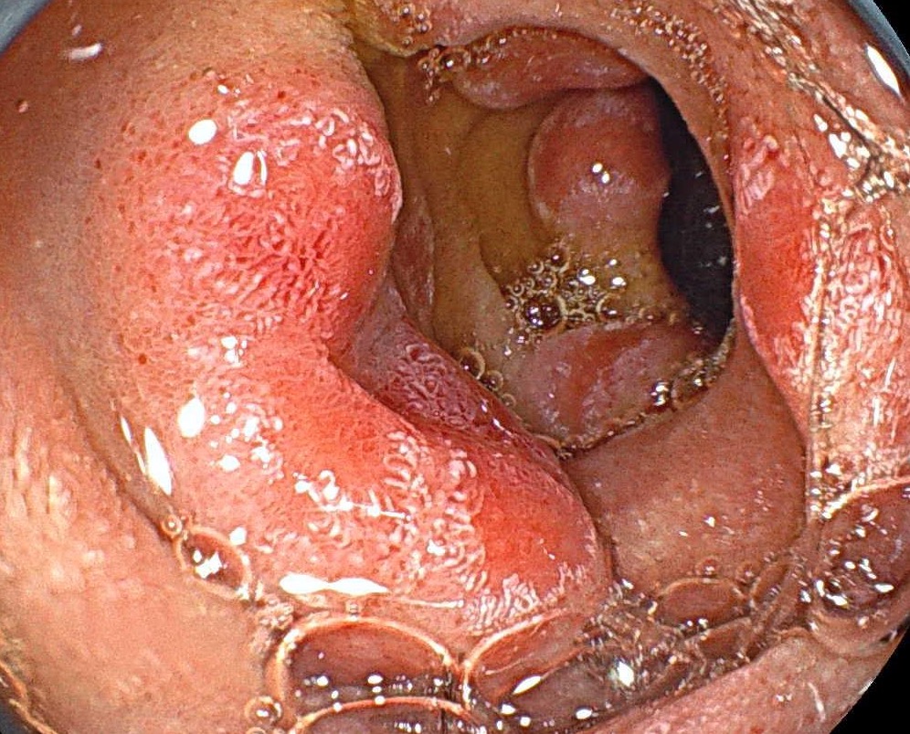
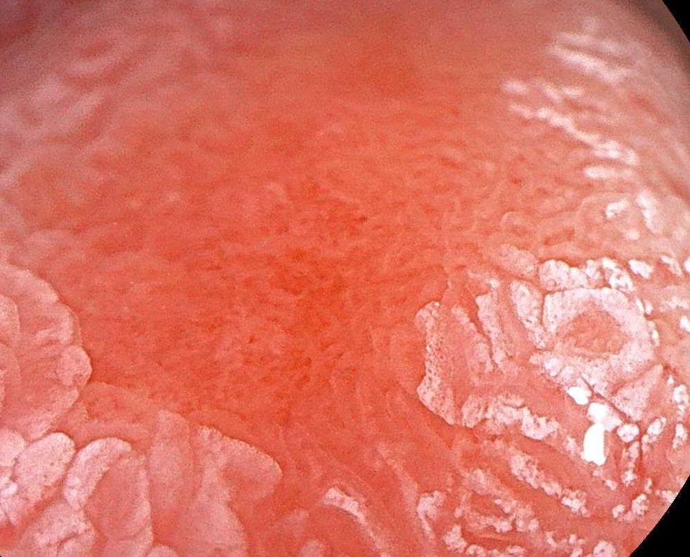
A 75-year-old man with no symptoms had a duodenal EMR 10 years ago, with normal follow-up biopsies. A recent surveillance endoscopy found an enlarged papilla, and he was referred for a second look. Tumor markers were negative and he had no diarrhea. The second look showed several raised, reddened nodular areas.
The first reply, 21 minutes later, read them as inflammatory. A senior member was less sure. Duodenal adenomas like to come back even after complete resection, so a recurrent adenoma or an adenocarcinoma had to be considered. The multifocal pattern made him think of lymphoma, such as follicular lymphoma. If the patient had cirrhosis, portal hypertensive duodenopathy with inflammatory polyps was possible. He had also recently seen blastomycosis look very similar in immunosuppressed patients. Another member suggested reactive lymphangiectasia.
A third member focused on the first lesion, which had a depression. The surrounding mucosa was not worrying, but the depression was. He would biopsy that lesion as though it were malignant, with at least eight biopsies from all sides. The other nodular areas could be peptic duodenitis, with lymphoma still on the list.
One member had seen a similar finding recently, where the biopsies showed only gastric metaplasia. Biopsies were taken in this case and the histology was still pending at the end of this window. We will report the result if it is shared.
Clinical Pearl: Duodenal adenomas often come back, even after complete resection. When new nodules appear years later, biopsy them thoroughly, take extra care with any depressed area, and keep lymphoma on the list when the change is multifocal.
3. A Migrated Hepaticogastrostomy Stent with No Wire Access
First reply in 7 minutes. 2 colleagues weighed in.
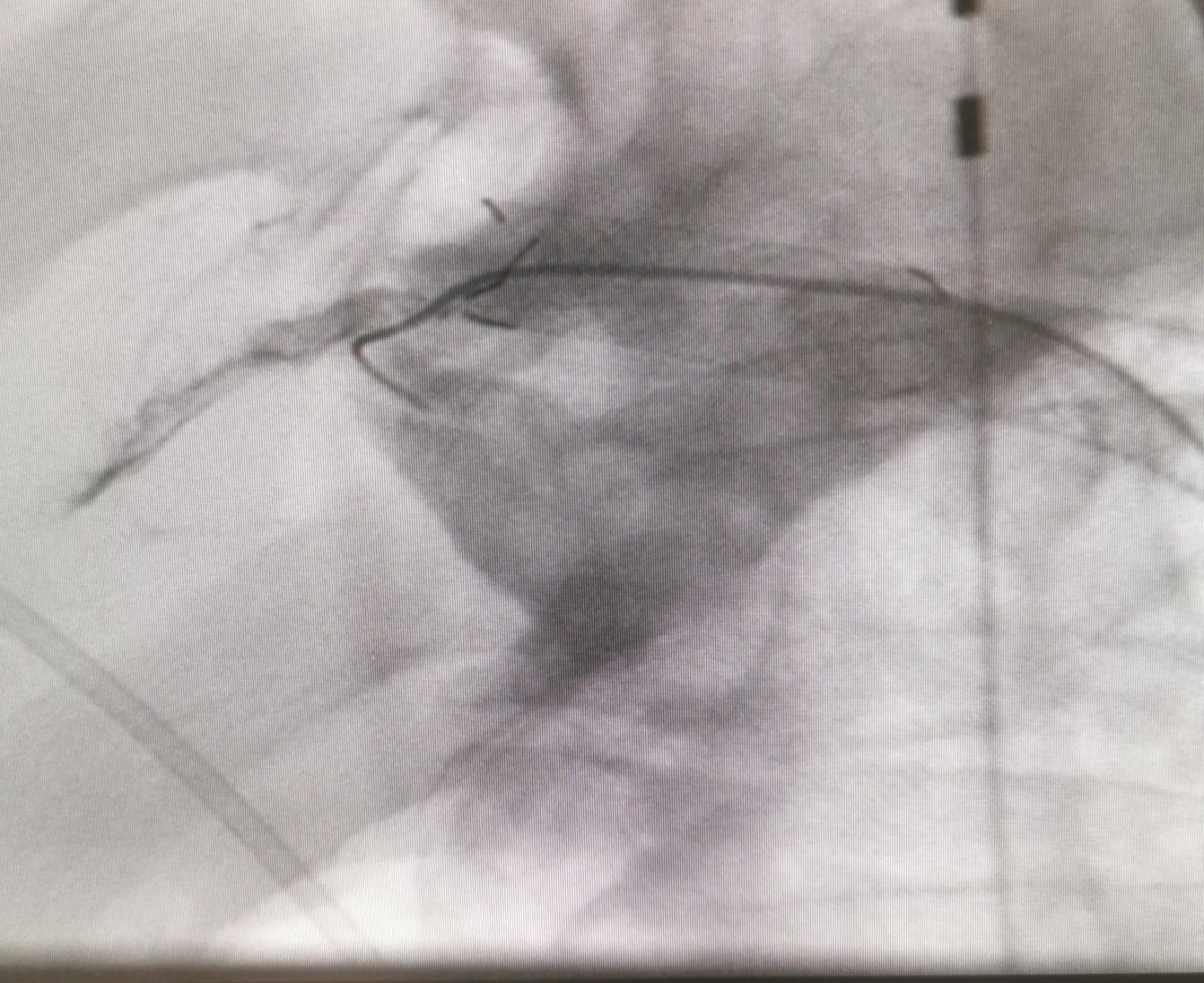
A patient with liver metastases from pancreatic cancer had a hepaticogastrostomy placed three weeks earlier. The stent had migrated. On CT it was still in the liver, but it no longer drained the left duct, and a guidewire would not pass into the biliary tree. ERCP was not an option, because the malignant duodenal stenosis was so tight that even contrast would not pass.
The first reply came 7 minutes later. The first questions were whether the stent was really still in the liver, since a stent in place should give access to the biliary radicals, and whether contrast had filled the ducts, because much of it seemed to lie outside. A plain CT was the suggested first step to confirm the stent still communicates with the liver. The presenter confirmed on CT that it did.
The suggested rescue was to go back through the same stent. Use a thinner wire, 0.018 or 0.025 inch, or an angled wire. Once a wire is across, dilate with a CRE balloon and place a new stent through the old one. The team planned to check which wires it had, and the senior endoscopist was considering replacing the transgastric stent.
Clinical Pearl: If a hepaticogastrostomy stent is still in the liver on CT but a wire will not pass, go back through the same stent with a thinner or angled wire, then dilate and place a new stent through the old one.
Follow-ups
From Issue 13 · Why a cholecystectomy clip can cause trouble 14 years later
In Issue 13, the presenter of the common hepatic duct stricture asked why a surgical clip took 14 years to cause a problem. The next day a member answered: clips can keep up inflammation around the duct for years, and can even migrate from outside the duct into it. He shared three published case reports of clip migration into the bile duct.
Quick Consultations
Bariatric · Gastric bypass for GERD and gastroparesis in one operation?
A patient with diabetes (HbA1c 9.0), a BMI of 25.6, grade B esophagitis, a hiatal hernia, and retained food despite fasting. One answer was yes, with conditions: 12 weeks of high-dose PPI has failed to heal the esophagitis on repeat endoscopy, nuclear imaging clearly shows gastroparesis, and the surgeon keeps the limb under 50 cm to avoid too much weight loss.
Bleeding · Refractory shock at night with no IR or surgical backup
A rural hospital with no IR, no surgical backup, and a tele-ICU: a patient in hemorrhagic shock on half-maximal norepinephrine plus vasopressin, with lactate not clearing. Scope at night, or transfer 30 minutes to a center with 24/7 IR and surgery? Most of the group said transfer. One member noted that patients die during endoscopy for upper GI bleeding when they are under-resuscitated or aspirate, but an unstable patient may not survive transfer either. The patient was transferred. CTA was negative, possibly falsely so on high-dose vasopressors.
Colon · A sigmoid ulcer after diarrhea that started before a trip
A 47-year-old had a month of non-bloody diarrhea that had almost settled by the time of colonoscopy, which showed sigmoid edema, hyperemia, and an ulcer. Stool tests so far were negative, and the diarrhea had worsened on a trip abroad. The group read it as the healing phase of an infectious colitis, such as E. coli or Shigella, which give linear deep ulcers in the acute phase. Not UC, and not NSAIDs. Schistosomiasis looks different, with yellowish nodules where the eggs are laid, and amoebic colitis usually gives volcano ulcers.
Liver · A 1.5 cm liver cyst, hepatomegaly, and a family history of cholangiocarcinoma
A 1.5 cm liver cyst with a 20 cm liver, normal liver tests, negative autoimmune, iron, and viral workup, a contrast allergy, and a prior sleeve gastrectomy. A cyst this small is most likely simple. Biliary cystadenoma usually shows multiple chambers, septations or wall nodules, and enhancing walls. With the usual causes ruled out, infiltrative or storage disease was raised for the hepatomegaly.
Pancreas · Probable main-duct IPMN in an 84-year-old: EUS if surgery is off the table?
An 84-year-old in good health had an incidental cystic lesion in the head of the pancreas, with normal CA 19-9, enzymes, and liver tests. On MRI the group read a probable main-duct IPMN, likely over 3 cm. One view: EUS, with a side-viewing look for a fish-mouth papilla, still settles the diagnosis and the level of risk even if no surgery is planned. The other: a Whipple carries a high mortality in octogenarians, so a test that will not change management may not be worth its small risk. Both sides agreed on a frank, shared decision with the patient.
Rectum · Rectal bleeding with prolapse and a probable solitary rectal ulcer
An elderly woman with rectal bleeding, prolapsing hemorrhoids, and what looked like a solitary rectal ulcer. The advice was referral to a colorectal surgeon, with biopsies to rule out malignancy, especially of a grey-white area seen in retroflexion. Biopsies were taken. A chronic anal fissure with prolapsing hypertrophic anal papillae was also suggested.
Small bowel · Pseudo-obstruction and pneumatosis in myeloma, with amyloid on biopsy
A 73-year-old with untreated myeloma had a laparotomy for suspected perforation that found nothing. A month later a CT was read as pneumatosis with cecal necrosis, although he felt almost well and his lactate stayed normal. Biopsies at EGD and colonoscopy showed amyloidosis. The group read the original obstruction as pseudo-obstruction from amyloid in the bowel wall. The pneumatosis may have come from the amyloid or from the laparotomy itself.
Upper GI · Aphthous ulcers from the esophagus to the bulb, calprotectin 450
Aphthous ulcers in the esophagus, stomach, and duodenal bulb, with a normal colon, a slightly red ileum, chronic diarrhea, and a fecal calprotectin of 450. H. pylori and NSAIDs were ruled out and tTG was negative. Upper GI Crohn's disease is on the list, but the group said aphthous ulcers and diarrhea are not enough to call it IBD. Next steps: MR enterography, biopsies for features of chronicity, duodenal biopsies for celiac disease, and CRP, albumin, B12, and iron studies.
Community Notes
- Hard decisions: Three questions one senior member asks himself before advising on a hard case. Will the result of this test change what we do? Do the benefits of treatment outweigh the risks, once all of the patient's other conditions are counted? And if this were my own mother or father, what would I recommend?
- Bariatric: One member's experience with five marginal ulcers at the gastrojejunal anastomosis after gastric bypass: three presented with bleeding and two with pain. All responded to a PPI and sucralfate. There is no set surveillance interval. He repeated endoscopy at three months, and most ulcers took 6 to 12 months to heal.
What’s Next
Every case in this digest came from real conversations in the EndoCollab private WhatsApp community, 650+ practicing endoscopists discussing cases daily from every continent. Lifetime members get permanent access to the group, plus the full EndoCollab case library.