COMMUNITY DIGEST
3 featured cases and 13 quick consultations from the EndoCollab WhatsApp community between July 18 – August 2, 2026. This period brought a dense mix of hepatology emergencies, biliary imaging, and practical endoscopy troubleshooting: a postpartum jaundice course that evolved into sepsis and DIC before recovery, an 89-year-old with double-duct sign on ERCP, and suspected chemical colitis from residual glutaraldehyde.
In this issue
- Postpartum Cholestatic Jaundice Evolving to Sepsis and DIC
- Double-Duct Sign and a Challenging Cholangiogram in an 89-Year-Old
- Segmental Colonic Injury: Is Residual Glutaraldehyde the Cause?
- Quick cases: Post-Banding Esophageal Variceal Rebleed; Esophageal Polyp: Papilloma vs Inflammatory Lesion; Isolated Sigmoid Dilation Without Clear Volvulus; Classic Celiac Pattern in the Second Duodenum; Hypopharyngeal Diverticulum; Undetectable Fecal Elastase and Chronic Diarrhea; Migrated CBD Stent: Straight vs Double-Pigtail Length; Biliary Stent Lodged in a Sigmoid Diverticulum; Acute HBV Flare in a Previously Immune-Tolerant Young Adult; Gastric Extrinsic Bulge: CT Before EUS; Post-Cholecystectomy Diarrhea and Bile Acid Binders; Late Post-Cholecystectomy CHD Stricture; Esophageal Granular Cell Tumor (Abrikossoff)
- Community notes: For subepithelial lesions, CT often shows extraluminal bulk…; For generalized abdominal lymphadenopathy, gastrohepatic or…; ACG guidance avoids routine urgent ERCP without cholangitis…; When TPMT is unavailable, one common practice pattern for az…; The group discussed ERBE effect, duration, and interval sett…
Featured Cases
1. Postpartum Cholestatic Jaundice Evolving to Sepsis and DIC
A 24-year-old primigravida underwent lower-segment cesarean section and was noted to have jaundice on day 0. Initial outside LFTs showed a cholestatic pattern (total bilirubin 8.1, direct 6.2, ALP 500, AST/ALT near 99). Same-day repeat labs showed rising bilirubin and ALP to 768, INR 1.5, creatinine 2.2, NAGMA on ABG, and one hypoglycemic episode (glucose 55). GCS remained 15/15. Ultrasound showed no choledocholithiasis and no fatty liver, with gallbladder wall edema, mild pancreatic head bulkiness, and minimal ascites. Pre-delivery LFTs one week earlier had been normal.
The community differential was broad: acute fatty liver of pregnancy (AFLP), shock liver, HELLP or pre-eclampsia spectrum, drug-induced liver injury, viral hepatitis, and less likely choledocholithiasis given absence of pain and normal ducts. Classic ischemic hepatitis usually produces much higher AST/ALT, which argued against pure shock liver. AFLP remained high on the list because of third-trimester and postpartum timing, hypoglycemia, and renal dysfunction. Members recommended serum bile acids, medication review, and close glucose and coagulation monitoring.
Within 24 hours the picture darkened: WBC rose from 20k to 44k with 89% neutrophils, platelets fell, APTT prolonged, D-dimer rose to about 7000, procalcitonin was 10, ammonia was 130, and MRCP showed no choledocholithiasis. The group reframed the case as evolving AFLP with sepsis and a DIC pattern, urged readiness for transfer to a transplant-capable center, early broad-spectrum antibiotics, albumin support, and avoidance of prophylactic clotting-factor transfusion unless bleeding or procedures required it.
Update: the patient improved. Bilirubin fell to 3, transaminases normalized, AKI recovered, and sepsis settled. Delivery plus aggressive supportive care reversed the course.
Clinical Pearl: Postpartum cholestatic jaundice with hypoglycemia, renal injury, and coagulopathy is AFLP until proven otherwise. Do not call the patient stable when labs show rising inflammatory markers, a DIC pattern, and ammonia elevation. Exclude retained products and bile duct obstruction, start sepsis care early, and involve a transplant center before encephalopathy closes the window.
2. Double-Duct Sign and a Challenging Cholangiogram in an 89-Year-Old
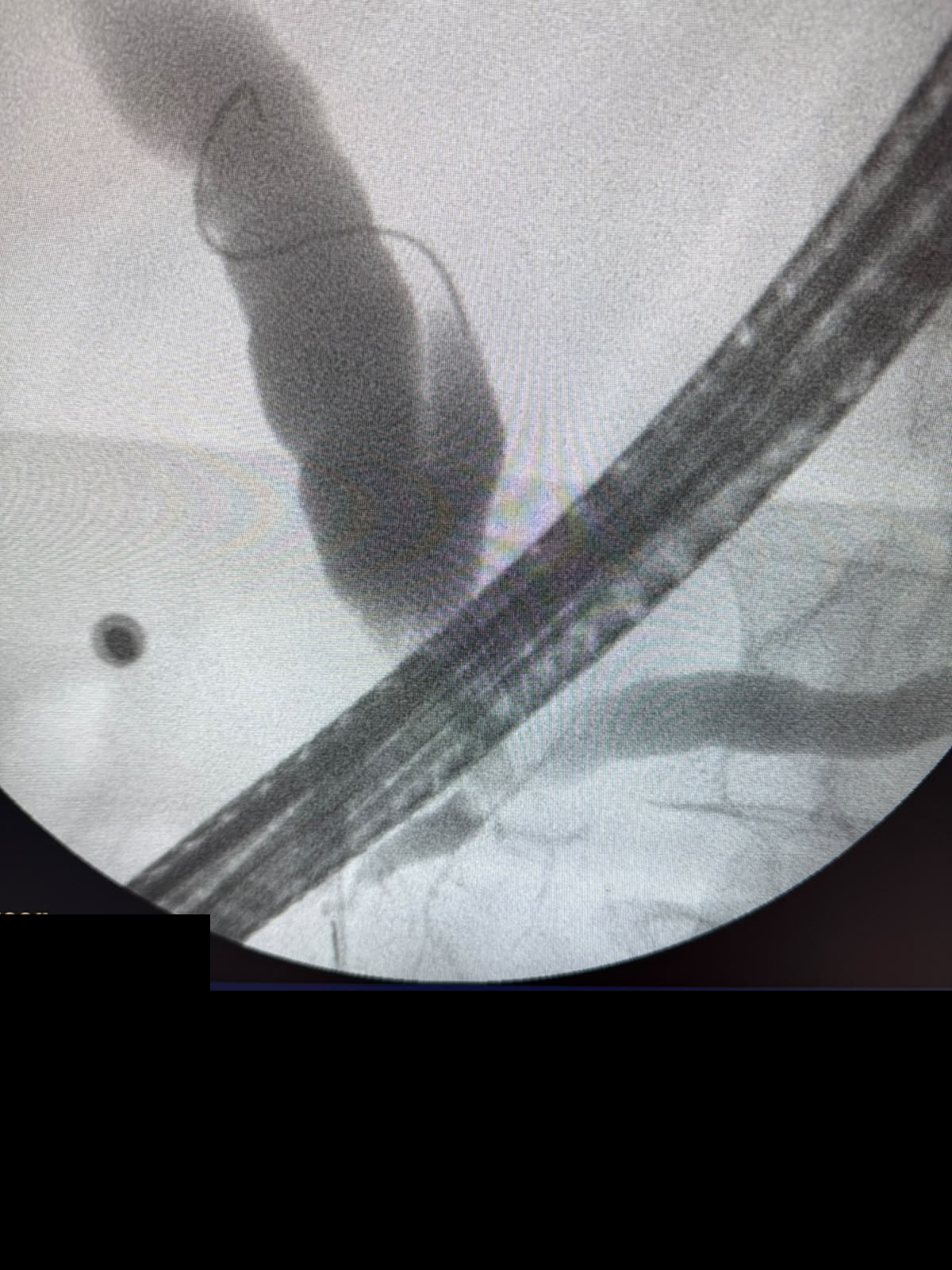
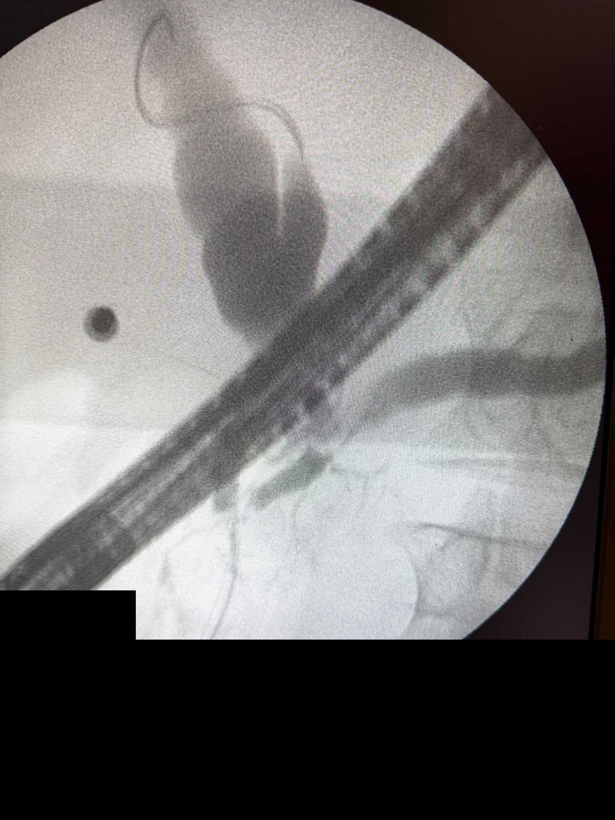
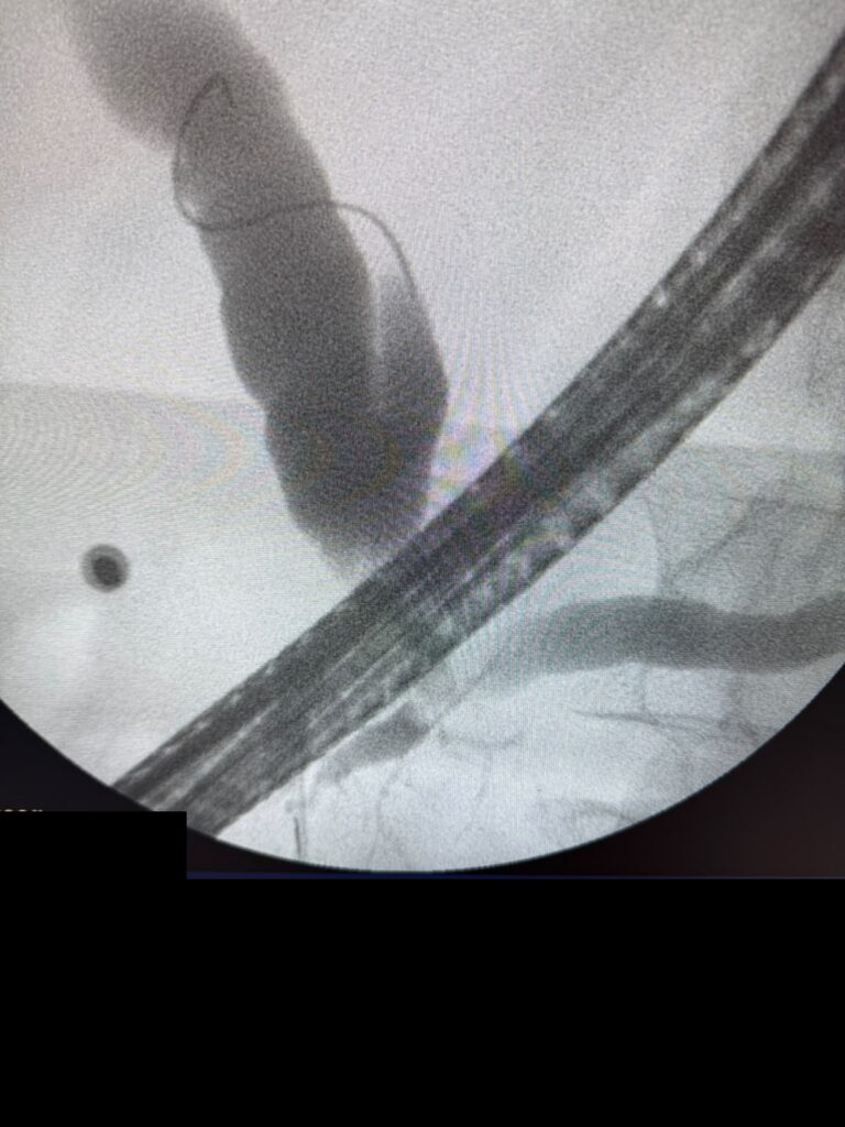
An 89-year-old presented with painless obstructive jaundice. MRI and MRCP showed simultaneous dilation of the common bile duct and main pancreatic duct (double-duct sign) without a definitive mass diagnosis. ERCP was performed for biliary drainage and further characterization.
Cholangiograms showed a sphincterotome in position, a guidewire course suggesting low or ectopic cystic duct insertion, and a dilated pancreatic duct. Members recommended contrast CT of the pancreatic head and careful interpretation of wire position relative to the cystic duct versus true ductal pathology.
Malignancy (periampullary or pancreatic head) remains the leading concern when double-duct sign accompanies painless jaundice in the elderly. IgG4-related disease stays on the broader stricture differential in other clinical contexts.
Clinical Pearl: Double-duct sign with painless jaundice in an older adult is cancer until excluded. Confirm wire and contrast anatomy before attributing every channel to pathology, and pair ERCP drainage with high-quality cross-sectional imaging of the pancreatic head.
3. Segmental Colonic Injury: Is Residual Glutaraldehyde the Cause?
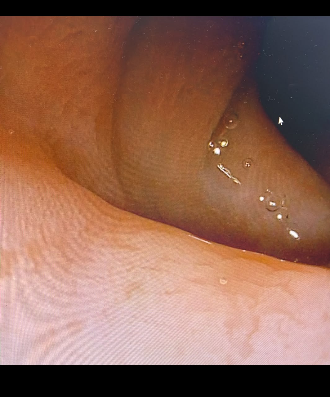
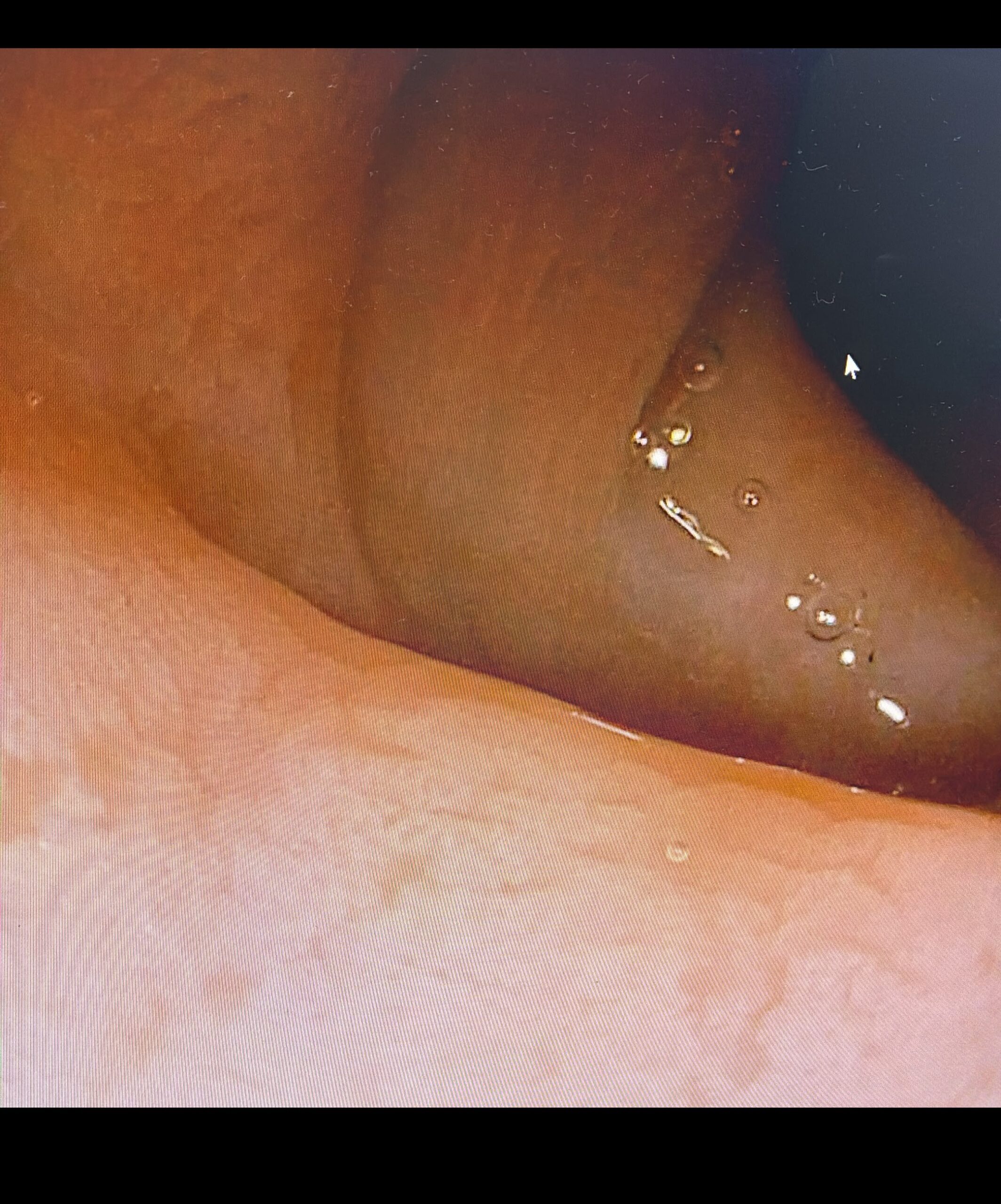
Endoscopy showed segmental mucosal changes in the colon, including the descending colon, with whitish debris and altered mucosa. The question was whether residual glutaraldehyde from endoscope disinfection could explain the findings.
The community agreed chemical colitis is a real possibility. Injury from inadequately rinsed glutaraldehyde is a classic iatrogenic pattern that can mimic ischemic or infectious colitis and is often segmental, corresponding to residual disinfectant contact.
Prevention centers on meticulous rinsing after high-level disinfection. New colitis after a recent procedure may be chemical rather than idiopathic inflammatory bowel disease.
Clinical Pearl: When segmental colitis appears shortly after endoscopy or on scopes processed with glutaraldehyde, consider chemical injury from residual disinfectant. Confirm rinse protocols before escalating IBD therapy.
Quick Consultations
Portal Hypertension · Post-Banding Esophageal Variceal Rebleed
A 58-year-old woman with prior repeated band ligation presented with massive hematemesis two weeks after the last banding session. Endoscopy showed spurting at one post-banding site and oozing at another; balloon tamponade was placed because vision was lost. Consensus: reattempt banding in an intubated ICU setting once the balloon is deflated, with vasoactive therapy running and rescue TIPS ready. A fully covered metal stent is preferred over prolonged balloon tamponade if endoscopic hemostasis fails. Tissue adhesive is more typical for fundal varices than esophageal varices in most practices.
Esophageal Pathology · Esophageal Polyp: Papilloma vs Inflammatory Lesion
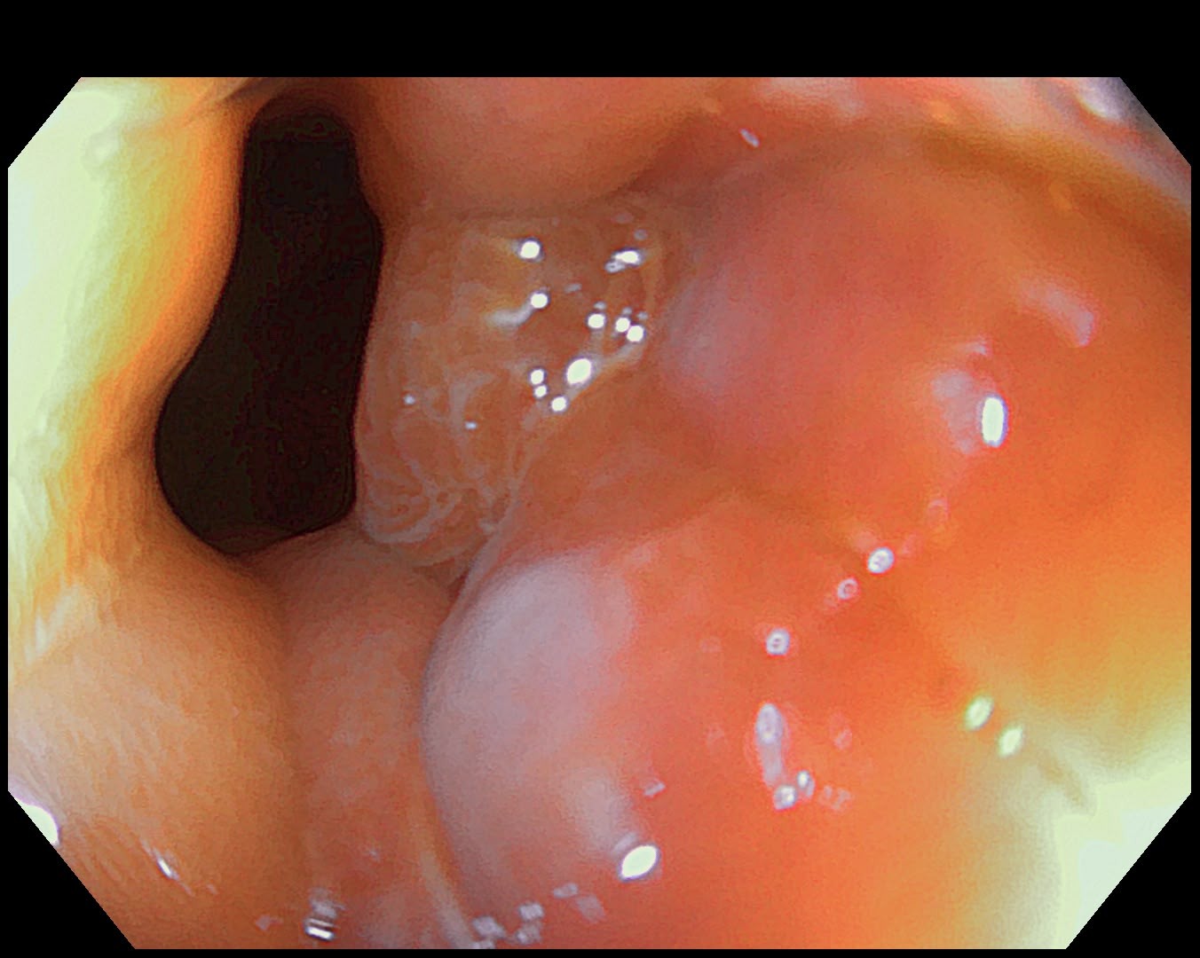
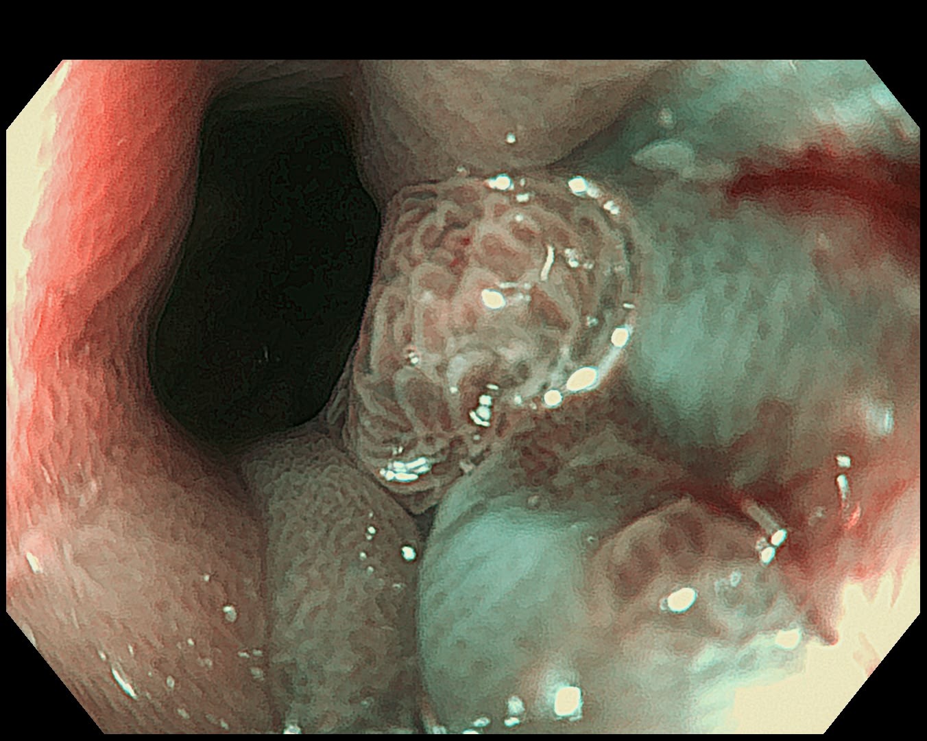
A polypoid esophageal lesion prompted discussion of squamous papilloma versus hyperplastic or inflammatory polyp, including GERD-related sentinel polyp at the cardia. Biopsy was obtained. The group favored benign epithelial polyp categories and stressed histology over visual overcall.
Colonic Motility · Isolated Sigmoid Dilation Without Clear Volvulus
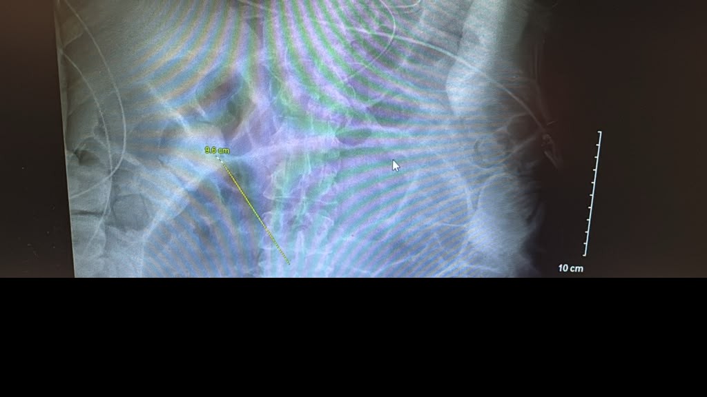
A 71-year-old ICU patient with COPD exacerbation and pneumonia requiring intubation had marked isolated sigmoid dilation. Contrast reached the rectum. CT lacked a clear transition point or whirl sign. The patient had stool output and no abdominal pain. Differential included Ogilvie-type colonic pseudo-obstruction, segmental megacolon, and occult volvulus. Plan discussed: flatus tube or endoscopic decompression, rule out C. difficile, and reserve neostigmine for true pseudo-obstruction without mechanical obstruction when clinical status allows.
Small Bowel · Classic Celiac Pattern in the Second Duodenum
Endoscopy for chronic diarrhea showed scalloping and mosaic pattern in D2. The group recognized textbook celiac disease endoscopically. Histology confirmation remains mandatory before lifelong gluten-free counseling.
Pharynx and Esophagus · Hypopharyngeal Diverticulum
A hypopharyngeal pouch seen on endoscopic evaluation was confirmed as consistent with hypopharyngeal (Zenker-type) diverticulum anatomy. Recognition matters for safe intubation and for planning definitive therapy when symptoms warrant.
Pancreatic Insufficiency · Undetectable Fecal Elastase and Chronic Diarrhea
A 77-year-old on hemodialysis with heavy alcohol history and up to 10 stools daily had negative stool microbiology, normal calprotectin, normal colonoscopy with biopsies, and fecal elastase under 1 ug/g on two samples, including a semi-formed sample on loperamide. The group favored possible exocrine pancreatic insufficiency from chronic pancreatitis even without prior acute pancreatitis. EUS or plain film can look for calcific disease. An empirical trial of pancreatic enzyme replacement (about 40,000 units with meals) is reasonable while workup continues. Watery stool can falsely lower elastase, but repeated undetectable values plus alcohol history support an enzyme trial.
ERCP · Migrated CBD Stent: Straight vs Double-Pigtail Length
After distal migration of a biliary stent outside the CBD, the group preferred a longer straight stent (for example 12 cm) over a double-pigtail of the same labeled length. Pigtail curls consume roughly 1.5 cm on each end and reduce effective bridging length across a stricture. An 18 cm double-pigtail would be needed if curls are required.
Foreign Body · Biliary Stent Lodged in a Sigmoid Diverticulum
An 87-year-old had a migrated biliary stent tracked on serial x-rays for two months without passage. Advice: bowel prep, same-day plain film, and endoscopic retrieval with rat-tooth forceps or snare if still present. Do not wait indefinitely once the stent is fixed in a tight diverticular segment.
Hepatology · Acute HBV Flare in a Previously Immune-Tolerant Young Adult
A 33-year-old with vertically acquired HBV, prior high viral load and normal LFTs years earlier, presented with ALT about 1900, AST about 1700, bilirubin about 100 umol/L, INR about 1.6, no encephalopathy, and improving labs. The group recommended ruling out HDV superinfection and other acute insults, and initiating antiviral therapy (entecavir or tenofovir) given severity even without encephalopathy.
Subepithelial Lesions · Gastric Extrinsic Bulge: CT Before EUS
An anterior-wall bulge below the fundus raised subepithelial lesion or GIST versus extrinsic compression from spleen, kidney, or other mass. With normal ultrasound, CT first was preferred to map the full extraluminal story. EUS follows when a wall-origin mass is confirmed or when small lesions are occult on CT.
Post-Surgical · Post-Cholecystectomy Diarrhea and Bile Acid Binders
Year-long diarrhea after laparoscopic cholecystectomy with normal colonoscopy. A brief cholestyramine trial was poorly tolerated. Colesevelam may be easier as tablets where available. Without SeHCAT, a properly dosed bile acid binder trial remains both diagnostic and therapeutic. Also consider SIBO, pancreatic insufficiency, and parasites before labeling bile acid diarrhea alone.
Biliary Strictures · Late Post-Cholecystectomy CHD Stricture
Remote cholecystectomy with early stenting, then years of intermittent jaundice treated with antibiotics, now bilirubin 6.4. Imaging suggested a CHD-region stricture. Need contrast-enhanced MRI (not MRCP alone), exclude cholangiocarcinoma and IgG4 disease, relieve obstruction with dilation and stenting or hepaticojejunostomy as anatomy dictates, and watch for secondary biliary cirrhosis.
Submucosal Tumors · Esophageal Granular Cell Tumor (Abrikossoff)
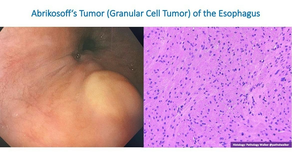
Discussion of a yellowish submucosal esophageal nodule as granular cell tumor (Vanek or Abrikossoff tumor) of Schwann cell origin. SOX10 and S100 are typically positive. Malignant transformation risk is low (about 2%). Unlike leiomyoma or lipoma, biopsies can often reach the diagnosis because these lesions sit near the mucosa and may erode.
Community Notes
- EUS Strategy: For subepithelial lesions, CT often shows extraluminal bulk that endoluminal views underestimate. EUS is essential for wall-layer origin and sampling when the mass is gastric, but CT first reduces surprises such as a renal mass indenting the stomach.
- Lymphoma Sampling: For generalized abdominal lymphadenopathy, gastrohepatic or periportal and portocaval nodes are accessible by EUS. FNA or FNB may still fail to subtype lymphoma depending on local pathology support, so plan for possible excisional biopsy.
- Gallstone Pancreatitis: ACG guidance avoids routine urgent ERCP without cholangitis or persistent obstruction. A 3 mm CBD stone with falling bilirubin can often be observed with serial labs and clinical monitoring.
- IBD Medications: When TPMT is unavailable, one common practice pattern for azathioprine titration is weekly dose steps with close CBC and liver enzyme monitoring.
- Electrosurgery: The group discussed ERBE effect, duration, and interval settings and principles of diathermy for matching energy to polyp type and resection technique.
What’s Next
Every case in this digest came from real conversations in the EndoCollab private WhatsApp community, 650+ practicing endoscopists discussing cases daily from every continent. Lifetime members get permanent access to the group, plus the full EndoCollab case library of 1,700+ teaching cases, technique videos, and references.