Barrett’s esophagus for endoscopists: Z line and palisade veins, Prague C/M length, Seattle biopsies, dysplasia detection, and ablation options. Full.
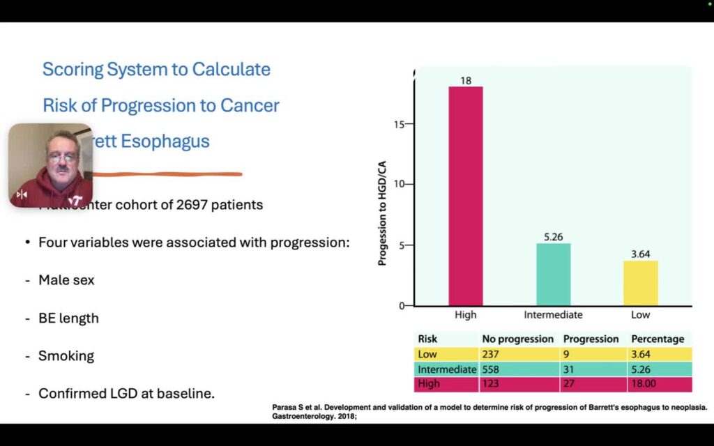
Barrett’s esophagus is a precancerous change of the distal esophagus that can progress to adenocarcinoma. This state-of-the-art overview frames why quality endoscopy matters: missed neoplasia is common, screening opportunities are often lost, and definitions still differ across societies (columnar metaplasia vs intestinal metaplasia with goblet cells). The lecture simplifies landmarks (Z line, GE junction, salmon-colored tongues, palisade veins), walks Prague C/M length, and ties Seattle four-quadrant sampling to the “Barrett’s archipelago” of patchy dysplasia. Risk context includes GERD-related injury, longer segment length, smoking, and confirmed low-grade dysplasia, with a reminder that many dysplasia labels need pathologist re-review. Practical detection emphasizes describing what you see (red, raised, nodular, ulcerated, irregular vessels on NBI) over forcing Paris polyp categories onto Barrett’s. Acetic acid for surface pattern, right-sided lesion bias, and a survey of eradication options (hybrid APC, Barrx/RFA, cryotherapy, endoscopic resection) round out the map for the full Barrett’s course.
Continue with EndoCollab membership.
Get this lecture, the rest of this course, and the full EndoCollab library.
Course: Barrett’s Esophagus · Browse all lessons in Barrett’s Esophagus
Continue reading with a membership
Get access to the full content, course videos, case library, and our private WhatsApp community.
Already a member? Log in
We protect your privacy. No spam. Unsubscribe anytime.