
By: By Klaus Mönkemüller, MD, PhD, FASGE, FJGES. Professor of Medicine, Virginia Tech Carilion School of Medicine, Virginia, USA
Hemoclips, originally designed for hemostasis, are versatile tools that have revolutionized various endoscopic procedures.
1. Understanding Hemoclips
Hemoclips are multifaceted tools that currently are used beyond their original purpose of hemostasis, and therefore we will refer to them as “clips”.These clips are now employed for closing perforations, post-resectiondefects, marking, and attaching devices such as stents and feeding tubes. For instance, in a procedure involving a gastric, esophageal or colon perforation, clips can effectively seal the defect, preventing further complications. Figure 1 shows several clinical applications of clips. A. Duodenal ulcer with visible vessel. B. Clip being pushed towards the lesion. C. Released clip closing the duodenal ulcer. D. Clipped Mallory Weiss tear. E. Large post-resection defect in the cecum. F. Complete prophylactic closure of the defect. G. Perforation of the cecum during endoscopic resection of fibrotic polyp. H. Complete closure of the defect with clips.
 1")
Hemoclips are indeed a testament to medical innovation.
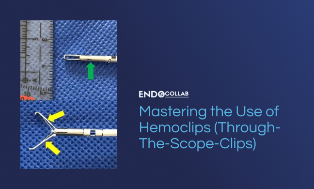
2. The Anatomy and Deployment Clips
Understanding the anatomy and mechanisms of action of clips is crucial to their effective use.
 2")
Clips consist of stem with two arms with distal teeth. The arms can vary in shape and number (yellow arrows). The length of the shaft or stem (green arrow) holding the arms can also differ, impacting their use in tight areas like small bowel strictures or esophageal stenosis. For example, a longer shaft in a small bowel stricture or esophagus could exacerbate the condition, as it would be rubbing the mucosa on the opposite site. Figure 1D shows a clip with “short” stem, and still, the stem’s end may still rub the opposite mucosa. A clip with long stem would have been inappropriate in this situation. Therefore, understanding the clip’s anatomy can guide its appropriate selection and use.
3. Advancing and Applying Clips
The advancement and application of through-the-scope clips require precision and care.
When first advancing the clip into the working channel of the scope, it’s crucial to hold close to the tip to avoid bending the arm. Once the clip has been pushed through the working channel and is at the tip of the scope, it should be gently pushed out, opened, and pulled back towards the scope (Figure 1B). For instance, when addressing a bleeding lesion, the clip should be open and advanced towards the lesion by moving the endoscope. At the final moment, once the clip is close to the target vessel or defect to be closed, the clip should be pushed out with the hand (Figure 1B), then closed and released by the assistant (Figure 1C).
Proper knowledge, handling and application of clips can significantly improve patient outcomes.
Remember, the mastery of clips lies in understanding their anatomy, their mechanics, knowing how to deploy them, and applying them with precision. With practice, you can harness the full potential of these versatile tools to improve patient care.

One Response
Very energetic article, I liked that a lot. Will there be a part 2?