A 3-minute guide to extreme ERCP: when the duodenoscope cannot pass the esophagus, build a bridge with an overtube and let a balloon scout the way.
Some ERCPs fail before the scope reaches the papilla. In this clip from his Extreme ERCP lecture, Klaus Mönkemüller defines a level V ERCP, then shows a patient with cholangitis whose esophagus was closed by cancer, radiation and a skin graft. The duodenoscope would not pass. Here is the route he built to the papilla.
Why the usual scale and the usual push do not help
The Schutz classification grades ERCP difficulty in four levels. Level 1 is deep cannulation or placing a stent. Level 4 is removal of intrahepatic stones, or ERCP after a Whipple procedure.
Device-assisted ERCP with single-balloon, double-balloon or spiral enteroscopes, ERCP with cholangioscopy, and rendezvous with percutaneous transhepatic cholangiodrainage (PTCD) do not fit that scale.
In this patient the barrier is the esophagus itself. It was impossible to advance the duodenoscope, so the access problem has to be solved before the papilla can be.
Step 1: Name the level
Klaus defines level V, or extreme, ERCP as the use of novel or modified scopes, tools, devices, accessories and techniques to solve or remediate complex primary, secondary or iatrogenically induced biliopancreatic tract disorders.
Video moment @ 0:05
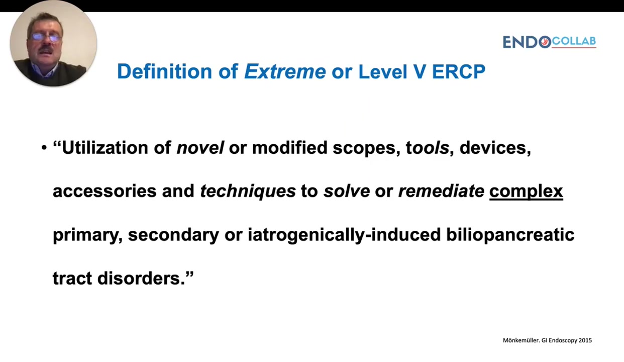
Significance: A level V case needs a plan for access and tools before it needs a plan for the papilla.
Step 2: See why the scope will not pass
The patient had cholangitis. He had a cancer, then radiation, then a skin graft placed into the esophagus. On endoscopy you can see the skin inside the esophagus, hairs from the graft, and his speech valve. It was impossible to advance the duodenoscope.
Video moment @ 1:11
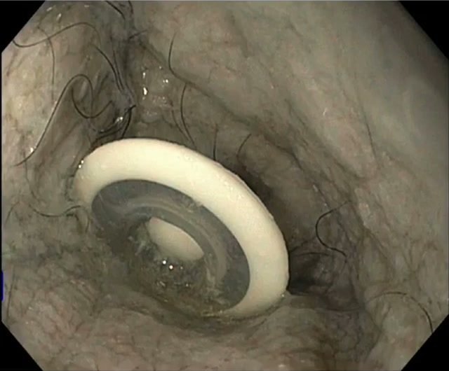
Significance: The obstacle is fixed anatomy. The plan has to change, not the push.
Step 3: Bridge it with an overtube
A Guardus (US Endoscopy) overtube was inserted into the upper esophagus. Then the duodenoscope, a side-viewing endoscope, was advanced through the overtube.
Video moment @ 1:19
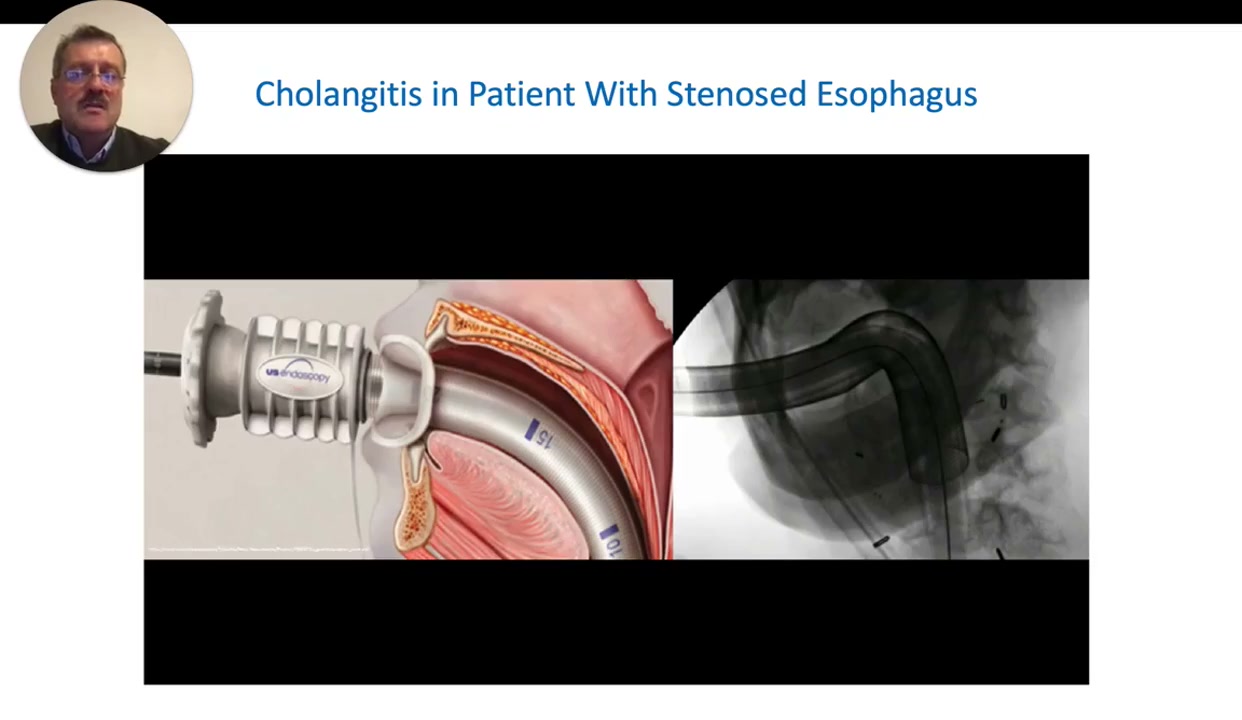
Significance: The overtube carries the duodenoscope past the graft and the speech valve, and keeps it stable.
The result you want in the note looks like this:
Access: Duodenoscope could not pass a stenosed esophagus (cancer, radiation, skin graft, speech valve). Guardus overtube placed in the upper esophagus. Duodenoscope advanced through the overtube.
Continue with a membership
Members watch the full 3-minute clip: the wire and CRE balloon that scout the stenosis, the ERCP, the same bridge in achalasia, and a paste-ready checklist.
Same format as our Prague classification guides: actionable steps you can use on the next case.
Already a member? Log in
Continue reading with a membership
Get access to the full content, course videos, case library, and our private WhatsApp community.
Already a member? Log in
We protect your privacy. No spam. Unsubscribe anytime.