It started during a colonoscopy. The clue was in her hair and nails.
A 2-minute case from my lecture on atypical polyps. Watch the case, make your call, then see the diagnosis.
1. The patient
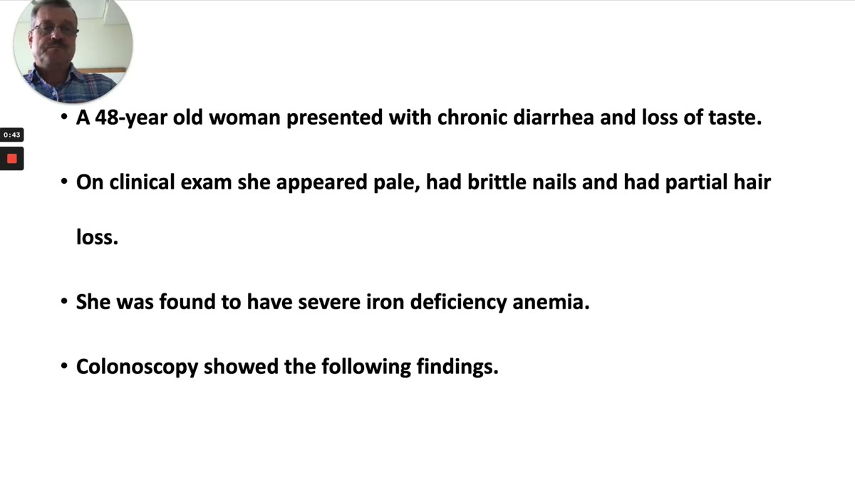
A 48-year-old woman presented with chronic diarrhea and loss of taste. On clinical exam she appeared pale, had brittle nails that were breaking down, and partial hair loss. Laboratory tests showed severe iron deficiency anemia.
2. Colonoscopy
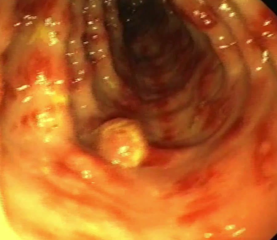
Colonoscopy showed a cecal polypoid lesion and many red streaks of the colonic mucosa that are raised and pseudopolypoid. There were many pseudopolyps and polyps, and some larger polyps too.
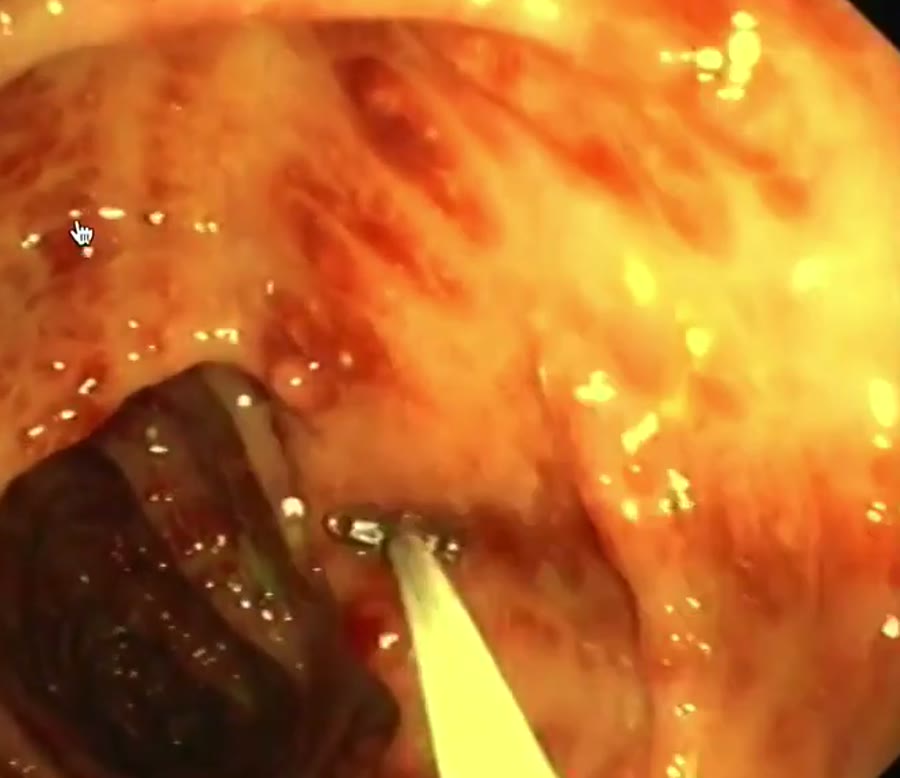
Some of these red streaks looked like a watermelon type colon, as we see with watermelon stomach. We obtained biopsies and also removed some polyps.
3. EGD
What impressed was a very thick gastric mucosa with pseudopolyps. In the duodenum there was patchy atrophy, and the villi were quite funny: like fingers, some longer than others, some on a very thin stalk. Some areas had no villi, so they looked like shaggy villi.
4. Histology
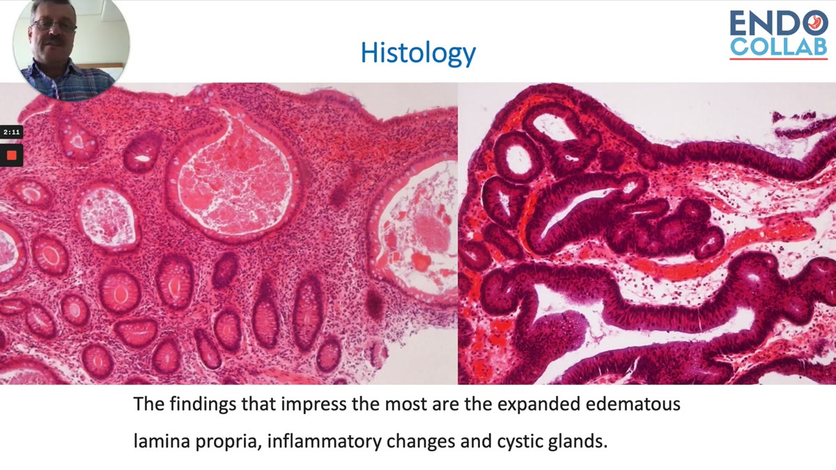
The findings that impressed most: an expanded, edematous lamina propria, inflammatory changes and cystic glands. The duodenal histology showed complete atrophy and lots of lymphocytic infiltrates.
5. What is your diagnosis?
Polyps and pseudopolyps in the colon, pseudopolyps in the stomach, an atrophic duodenum, iron deficiency, and a patient losing her hair and nails. Make your call before you read on.
What did colonoscopy show in this case?
A cecal polypoid lesion, many pseudopolyps and polyps, some larger, and many raised red streaks of the colonic mucosa. Some of the streaks looked like a watermelon colon, the pattern we know from watermelon stomach. We took biopsies and removed some polyps.
What did the stomach and duodenum show?
EGD showed a very thick gastric mucosa with pseudopolyps. The duodenum had patchy atrophy and odd villi: finger-like, some longer than others, some on a very thin stalk, and areas with no villi at all. Duodenal histology showed complete atrophy and many lymphocytic infiltrates.
The answer is at 2:16 in the full clip
The name, the clues that should tip you off, the cancer risk, and the treatment.
Members watch the full lecture and read the answer below, with the checklist for the next patient. Plus every other course video and case.
See the diagnosis: $29.99 a month
Cancel anytime. Or save with the yearly plan.
Not for you? Full refund within 30 days, for any reason.
Already a member? Log in and the full lecture plays.
Continue reading with a membership
Get access to the full content, course videos and the case library. Lifetime members also get our private WhatsApp group as a free bonus.
Already a member? Log in
We protect your privacy. No spam. Unsubscribe anytime.