Case Report


 Yuli Toledo, MD; Rafael Orellana, MD; Abel Sanchez, MD
Department of Gastroenterology, Hospital Roosevelt, Guatemala
Figure 1 · Spurting D2 Dieulafoy, one clip


Experienced teaching points
Clinical Pearls
01A Dieulafoy’s lesion is a dilated, tortuous arteriole that erodes through a small mucosal defect. There is usually no ulcer around it.
02When the vessel is visible and spurting, a single well-placed clip can stop the bleed immediately.
03Mechanical compression is a first-line option here. Injection or thermal therapy can wait if the clip already closed the vessel.
04These lesions bleed hard and can be missed. If the first look is dry, go back during the next bleed.
Get cases like this Saturday mornings. Free on The Practicing Endoscopist.
Final Diagnosis
Duodenal Dieulafoy’s lesion with acute gastrointestinal bleeding, treated with a single endoscopic clip.
Clinical History
A 37-year-old woman with chronic kidney disease and diabetes presented with one day of hematochezia. She was diaphoretic and hypertensive. Emergency endoscopy was performed for acute gastrointestinal bleeding.
Endoscopic Findings
- A spurting bleeding point in the second portion of the duodenum.
- A protruding vessel without surrounding ulceration, the typical Dieulafoy’s pattern.
Endoscopic Technique
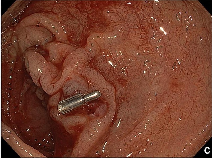
One hemostatic clip was placed on the spurting vessel in D2. Hemostasis was immediate. No further therapy was needed, and there were no complications.
Discussion
Mechanism. A Dieulafoy’s lesion is an abnormally large submucosal arteriole that erodes through a pinpoint mucosal defect. The surrounding mucosa is usually intact. That is why the bleed can look like nothing until the vessel jets, and why it can recur if the first exam is done between bleeds.
What the scope shows. Panel A is the emergency: active spurting in D2. Panel B is the diagnostic still: a nipple-like vessel with no ulcer crater. Panel C is the treatment: one through-the-scope clip, vessel closed, field dry.
Why it matters clinically. Injection and thermal therapy still have a role in GI bleeding. For a visible, spurting Dieulafoy, a clip gives mechanical occlusion you can see. Place it while the vessel is in view.
References
- Veldhuyzen van Zanten SJO, Bartelsman JF, Schipper ME, Tytgat GN. Endoscopic sclerosis of a Dieulafoy’s lesion. Endoscopy. 1986;18(5):211-213.
- Lee HY, Lim PN, Tham DK, et al. Endoscopic treatment of Dieulafoy lesions. Clin Endosc. 2011;44(4):279-281.
- Loffeld RJLF. Dieulafoy’s lesion: endoscopic diagnosis and treatment. Dig Liver Dis. 2011;43(2):103-105.
For your teaching file
Save this case as a PDF
Opens the companion page on The Practicing Endoscopist.