Case Report
 Klaus Mönkemüller, MD, PhD, FASGE, FESGE, FJGES
Carilion Clinic / Virginia Tech Carilion School of Medicine, Roanoke, VA, USA
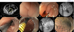
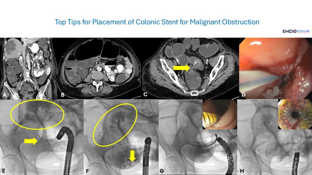
Figure 1 · Endoscopic and fluoroscopic steps

Experienced teaching points
Clinical Pearls
01Use a therapeutic endoscope with a working channel of at least 3.7 mm. Smaller channels often cannot accept the SEMS delivery system.
02Combined endoscopic and fluoroscopic guidance is essential for wire crossing, deployment monitoring, and confirming expansion.
03The balloon pullback technique estimates stricture length quickly: inflate distal to the stenosis and pull back until resistance.
04During deployment, pull back the scope and delivery catheter together. A distally expanding stent can shoot proximally if you stay still.
Get cases like this every other Saturday. Free on The Practicing Endoscopist.
Final Diagnosis
Malignant colonic obstruction due to metastatic colon cancer, treated with endoscopic placement of a self-expanding metal stent.
Clinical History
A 70-year-old woman presented with acute colonic obstruction in the setting of metastatic colon cancer. CT showed a large metastasis with distended large and small bowel loops. The indication for endoscopy was decompressing self-expanding metal stent placement.
Endoscopic Findings
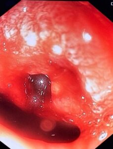
- Tight stenosis in the distal sigmoid colon on endoscopy.
- CT confirmation of obstruction with dilated proximal bowel loops and a large metastasis.
Endoscopic Technique
- Therapeutic gastroscope with a 3.7 mm working channel.
- Biliary balloon catheter and 0.035-inch J-tip guidewire advanced across the stenosis under combined endoscopic and fluoroscopic guidance.
- Contrast through the catheter confirmed position in the dilated proximal colon, then deep wire advancement for safe stent delivery length.
- Balloon inflated distal to the stricture and pulled back until resistance to estimate length and choose stent size.
- SEMS advanced over the wire. Proximal flange released under endoscopic view while gently pulling back both the endoscope and delivery catheter to prevent proximal stent shoot-up.
- Delivery catheter marking monitored for proximal positioning. Immediate endoscopic and fluoroscopic confirmation of expansion and decompression.
Discussion
Why SEMS here. Malignant colonic obstruction often needs urgent decompression. In advanced disease or high surgical risk, SEMS provides palliation or a bridge to surgery without an emergent resection or colostomy.
Equipment and imaging. Channel size is not a detail. Pediatric or thin channels under about 3.2 mm commonly fail to accept the delivery system. Dual guidance keeps the wire honest and shows deployment in real time.
Length and deployment control. Contrast plus balloon pullback improves stent sizing and coverage. The synchronized scope-and-catheter pull-back is the maneuver that keeps a distally expanding stent from migrating proximally. Full expansion can continue over hours to days; immediate balloon dilation is often unnecessary unless the lumen stays critically narrow.
References
- Small AJ, Coelho-Prabhu N, Lieberman DA, et al. American Society for Gastrointestinal Endoscopy guideline on the role of endoscopy in the management of colorectal cancer. Gastrointest Endosc. 2017;85(4):689-701.
- Van Hooft JE, van Halsema EE, Vanbiervliet G, et al. Self-expandable metal stents for the palliation of malignant colonic obstruction: updated recommendations from the European Society of Gastrointestinal Endoscopy. Endoscopy. 2014;46(12):1070-1081.
- Spinelli A, Galasso E, Contino G, et al. Endoscopic stenting for malignant colorectal obstruction: A systematic review and meta-analysis of outcomes in 2200 patients. Dig Liver Dis. 2018;50(1):11-18.
For your teaching file
Save this case as a PDF
Opens the companion page on The Practicing Endoscopist.