Technique Case

 Diana L. Dougherty, MD; Klaus Mönkemüller, MD, PhD, FASGE, FESGE, FJGES
Virginia Tech Carilion School of Medicine, Roanoke, Virginia, USA
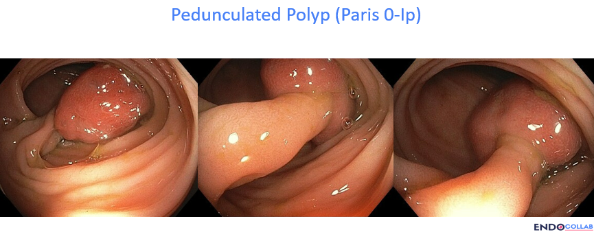
A 60-year-old man with a history of hypertension, coronary artery disease, and atrial fibrillation on Eliquis (stopped 2 days before colonoscopy) was found to have a large pedunculated polyp in the sigmoid colon.
Figure 1 · Pedunculated polyp (Paris 0-Ip)

Before you read on
How would you resect this polyp?
- aJust snare
- bPre-injection and then snare
- cSnare and then clip
- dPre-clipping and then snare
- eInject, clip, and snare
- fInject, snare, and then clip the stump
- gPlace an endoloop and then snare
- hSnare and then place an endoloop
- iUnderwater snare resection
- jOther
Answer
All options are correct. However, when dealing with pedunculated polyps, consider that there is always a large feeding vessel going through the pedicle. Clues to the presence of larger vessels are a) a large polyp head (needs more blood flow for "nutrition"), b) advanced adenomatous features (which correlate with neoplasia and angioneogenesis, i.e., more vessels/microcirculation in the polyp head), and c) thickness of the stalk or pedicle.
Many endoscopists prefer to cut first and then provide prophylactic or active hemostasis. However, this option may be risky, as post-resection bleeding may be brisk, especially in patients on anticoagulation or those who have bleeding diathesis or dyscrasias. Thus, I prefer to always perform the prophylactic hemostasis BEFORE polyp resection.
The most practical way to provide hemostasis is using hemoclips. If available, endoloops are quite useful and safe. Nevertheless, placing loops in angled positions such as the sigmoid colon can be challenging. Using a little pre-injection of saline or saline with epinephrine 1:20,000 into the base can also help create a temporary tamponade and decrease blood flow to the polyp head. However, sometimes injection can deform the stalk and make subsequent resection more challenging. Using underwater or saline-submerged resection has the advantages of a) less tissue damage during cutting and b) dilution of any post-polypectomy bleeding, with improvement of the visual field.
Get cases like this Saturday mornings. Free on The Practicing Endoscopist.
Figure 2 · Pre-clipping and the feeding vessel

In this case we opted for pre-clipping using two clips, followed by hot snare resection above the clips, proximal to the polyp head. Look at the stalk (yellow circle). Wow, that was a big feeding vessel! (yellow arrows). The choice of pre-clipping was great!
For your teaching file
Save this case as a PDF
Opens the companion page on The Practicing Endoscopist.