Case Report

 Byron Isaac Lewin Quijada, MD, Jose Gabriel Ruiz Rodriguez, MD, and Abel Alberto Sánchez Orozco, MD
Gastroenterology and Digestive Endoscopy Unit, Roosevelt Hospital, Guatemala City, Guatemala
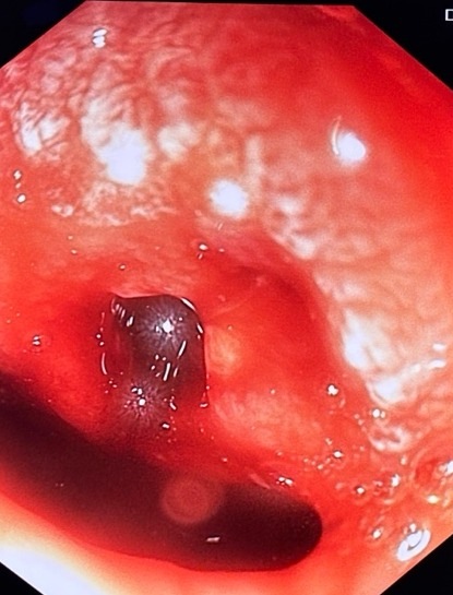
Figure 1 · Endoscopy, CT, and embolization


Experienced teaching points
Clinical Pearls
01After recent upper abdominal surgery, unexplained massive upper GI bleeding belongs on the portoenteric-fistula list, not only peptic ulcer.
02A clot-filled pylorus is not a finished exam. Evacuate the clot and take a second look at the duodenal bulb.
03Endoscopy shows the orifice. Contrast-enhanced CT maps the portal vein pseudoaneurysm and the adjacent hematic collection.
04If coils, hemostatic powder, and an over-the-scope clip are not on the shelf, transfer for transcatheter embolization instead of waiting.
Get cases like this every other Saturday. Free on The Practicing Endoscopist.
Final Diagnosis
Portal vein pseudoaneurysm-duodenal fistula after sequential abdominal surgery, treated with transcatheter coil embolization.
Clinical History
A 57-year-old man with no known chronic medical conditions presented with one day of hematemesis. He had undergone laparoscopic cholecystectomy, then exploratory laparotomy for perforated appendicitis ten days later. Nausea and epigastric pain preceded the bleed.
Admission hemoglobin was 6.4 g/dL. White blood cell count 9.60 K/µL, platelets 388 K/µL, prothrombin time 15.1 seconds (INR 1.39), and partial thromboplastin time 30.4 seconds. He was referred for emergent upper endoscopy.
Endoscopic Findings
- The first EGD showed a large adherent clot occupying the pyloric channel. The underlying lesion could not be characterized.
- Second-look endoscopy after clot evacuation identified a fistulous orifice in the duodenal bulb.
- There was no active bleeding at the moment of inspection.
Endoscopic Technique
Emergent EGD was performed for hematemesis and severe anemia. After clot evacuation, the bulb was re-inspected. Dedicated endoscopic hemostatic tools (coils, hemostatic powder or sponge, over-the-scope clip) were not available, so endoscopic closure was not attempted.
Contrast-enhanced CT demonstrated a pseudoaneurysm of the anterior branch of the right portal vein, an adjacent hematic collection along the wall of the second portion of the duodenum, and pneumoperitoneum. The patient was transferred for transcatheter coil embolization. Follow-up CT showed complete resolution of the pseudoaneurysm. There was no recurrent hematemesis or melena. Later hemoglobin was 8.8 g/dL.
Discussion
Mechanism. A portoenteric fistula forms when a portal vein pseudoaneurysm, produced by local inflammation, surgical dissection, or erosive injury, ruptures into adjacent bowel. The duodenum is the usual target because it sits against the portal triad in the hepatoduodenal ligament. Most published cases follow penetrating peptic ulcer, biliary or pancreatic surgery, chronic pancreatitis with a pseudocyst, or pancreaticoduodenectomy.
What the scope and CT show. Endoscopy may show only a hole, an ulcer look-alike, or a clot. Here the first look stopped at a pyloric clot. The second look found the orifice in the bulb. CT then did the vascular work: named the anterior branch of the right portal vein, mapped the hematic collection along D2, and documented free air. Because an isolated endoscopic hole can be mistaken for a simple ulcer, recent upper abdominal surgery plus unexplained hemorrhage is the cue to keep looking.
Why it matters. Reported mortality is high when the diagnosis is late. Options include endoscopic hemostasis (including EUS-guided coil embolization), transcatheter or transhepatic portal venous embolization, stent-graft placement, and surgery. The available toolkit decides the first move. Missing coils, powder, and OTSC made prompt IR transfer the correct first-line therapy, and it closed the pseudoaneurysm without recurrent bleeding.
References
- Burke CT, Park J. Portal vein pseudoaneurysm with portoenteric fistula: an unusual cause for massive gastrointestinal hemorrhage. Semin Intervent Radiol. 2007;24(3):341-345.
- Chantarojanasiri T, Sirinawasatien A, Bunchorntavakul C, Siripun A, Treepongkaruna SA, Ratanachu-Ek T. Endoscopic ultrasound-guided vascular therapy for portoduodenal fistula. Clin Endosc. 2020;53(6):750-753.
- Soares MA, Wanless IR, Ambus U, et al. Fistula between duodenum and portal vein caused by peptic ulcer disease and complicated by hemorrhage and portal vein thrombosis. Am J Gastroenterol. 1996;91:1462-1463.
- Lim SG, Park SE, Nam IC, et al. Large gastroduodenal artery pseudoaneurysm, arterioportal fistula and portal vein stenosis in chronic pancreatitis treated using combined transarterial embolization and transportal stenting: a case report. Medicine (Baltimore). 2022;101(52):e32593.
For your teaching file
Save this case as a PDF
Opens the companion page on The Practicing Endoscopist.