Case Report
 Geanfranco Vega, MD; Andrés Gutiérrez, MD
Universidad Mayor de San Marcos, Lima, Peru; Universidad de La República, Montevideo, Uruguay
Final Diagnosis
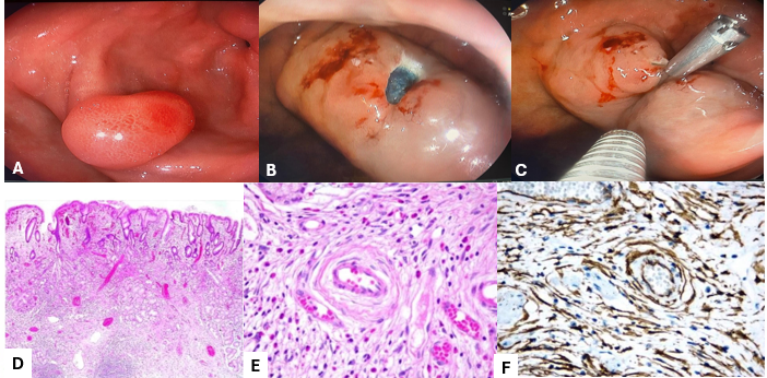
Gastric inflammatory fibroid polyp (Vanek tumor), resected via endoscopic mucosal resection.
Figure 1 · Vanek tumor of the stomach

Patient Demographics
A 50-year-old female patient.
Clinical History
The patient presented with epigastric abdominal pain. An upper endoscopy was performed to investigate the symptoms.
Endoscopic Findings
Endoscopy revealed a sessile polyp in the antrum, approximately 18 to 20 mm in size.
Endoscopic Technique
- The lesion was injected with a mixture of saline and blue dye.
- Endoscopic mucosal resection was performed.
- The post-resection defect was closed with a clip.
Discussion
Inflammatory fibroid polyps, also known as Vanek tumors, can occur throughout the gastrointestinal tract, most commonly in the stomach (approximately two-thirds of cases), followed by the ileum.
Initially considered inflammatory lesions, these are now recognized as true benign tumors due to an activating mutation of the PDGFRA gene (platelet-derived growth factor receptor alpha).
Microscopically, these polyps exhibit a classic onion-skin pattern, characterized by spindle cells whorling around prominent small blood vessels, alongside abundant eosinophils.
Most Vanek tumors are asymptomatic. However, some can lead to symptoms such as chronic abdominal pain, dyspepsia, bleeding, iron deficiency anemia, and, if in the small bowel, intussusception and bowel obstruction.
Asymptomatic and small polyps do not require specific therapy. However, polyps larger than 15-20 mm in the stomach or larger than 10 mm in the bowel warrant removal.
Endoscopic resection is the preferred therapeutic approach, as demonstrated in this case.
Immunohistochemistry was crucial for diagnosis, showing positivity for CD34 and negativity for CD117 (c-KIT negative) and DOG1, effectively ruling out a gastrointestinal stromal tumor (GIST).
Key Learning Points
01Vanek tumors are benign gastrointestinal stromal lesions linked to PDGFRA gene mutations.
02Gastric Vanek tumors over 1.5 cm typically warrant endoscopic resection.
03Immunohistochemistry is essential to differentiate Vanek tumors from GIST, with CD34 positivity and CD117/DOG1 negativity characteristic of Vanek tumors.
04Endoscopic mucosal resection is a safe and effective treatment for gastric Vanek tumors.
05The classic microscopic finding is an onion-skin pattern of spindle cells around blood vessels with eosinophilic infiltration.
References
- Vanek J. Gastric submucosal granuloma with eosinophilic infiltration. Am J Pathol. 1949;25(3):397-411.
- Daum O, Hatlova J, Mandys V, et al. Comparison of morphological, immunohistochemical, and molecular genetic features of inflammatory fibroid polyps (Vanek’s tumors). Virchows Arch. 2010;456(5):491-497.
For your teaching file
Save this case as a PDF
Opens the companion page on The Practicing Endoscopist.