
TECHNIQUE ARTICLE
Klaus Mönkemüller, MD, PhD, FASGE, FESGE, FJGES
Professor of Medicine, Department of Gastroenterology, Carilion Memorial Hospital, Virginia Tech Carilion School of Medicine, Roanoke, USA
Correspondence: Klaus Mönkemüller, MD, PhD. Department of Gastroenterology, Carilion Memorial Hospital, Virginia Tech Carilion School of Medicine, Roanoke, VA, USA
Abstract
Background: Patients with celiac disease who remain symptomatic or worsen despite a gluten-free diet need a structured reassessment. Most will have ongoing gluten exposure, an alternate diagnosis, or a functional overlap syndrome, but a smaller group has complicated celiac disease, including refractory celiac disease, ulcerative jejunoileitis, enteropathy-associated T-cell lymphoma, or small bowel adenocarcinoma.
Approach: This review summarizes when to move beyond repeat serology and duodenal biopsies to capsule endoscopy, deep enteroscopy, and radiologic imaging. Endoscopic warning findings include ulcerations, polypoid or tumor-like lesions, and raised discoid lesions. Radiologic warning findings include reversed jejunoileal fold pattern, small bowel dilatation, bowel wall thickening, cavitating mesenteric lymphadenopathy, mesenteric hypervascularity, and splenic atrophy.
Conclusion: Endoscopic and radiologic testing should be escalated when symptoms persist or worsen, when alarm features are present, or when refractory celiac disease or malignant complications are suspected. The goal is to identify treatable complications before they present as obstruction, bleeding, severe malnutrition, or advanced lymphoma.
Keywords: unresponsive celiac disease; non-responsive celiac disease; refractory celiac disease; capsule endoscopy; deep enteroscopy; MR enterography; enteropathy-associated T-cell lymphoma; small bowel adenocarcinoma; hyposplenism; splenic atrophy
Key Clinical Takeaways
- Persistent or worsening symptoms in a patient with celiac disease should first trigger a structured reassessment of the original diagnosis, gluten exposure, adherence, and alternate causes of symptoms.
- Escalate to capsule endoscopy, deep enteroscopy, CT enterography, MR enterography, or small bowel follow-through/enteroclysis when symptoms persist, alarm features appear, or complicated celiac disease is suspected.
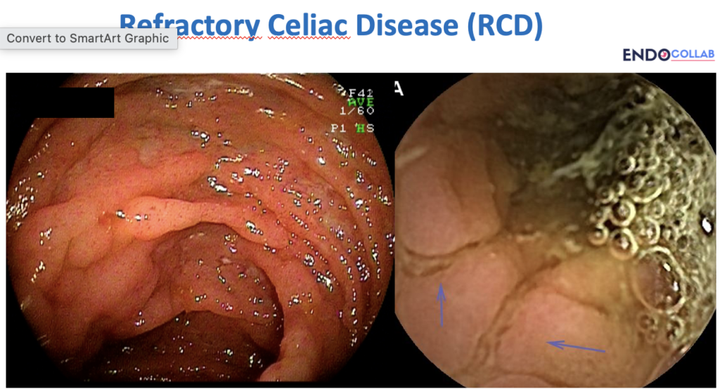
- Endoscopic warning signs include ulcerations, polypoid or tumor-like lesions, and raised discoid lesions in the small bowel.
- Radiologic warning signs include reversed fold pattern, small bowel dilatation, bowel wall thickening, cavitating mesenteric lymphadenopathy, mesenteric hypervascularity, and splenic atrophy.
- Splenic atrophy and functional hyposplenism matter clinically because they increase susceptibility to serious infection, especially pneumococcal infection. Vaccination status should be reviewed.
Clinical Problem
A patient with celiac disease who does not improve as expected is not a routine follow-up problem. Persistent diarrhea, abdominal pain, weight loss, anemia, fever, night sweats, obstructive symptoms, or worsening nutritional status should change the clinical frame. The question is no longer only whether the gluten-free diet is working. The question is whether the patient has non-responsive celiac disease, refractory celiac disease, or a complication such as ulcerative jejunoileitis, enteropathy-associated T-cell lymphoma, or small bowel adenocarcinoma.
Most patients with persistent symptoms will not have lymphoma. Gluten exposure, irritable bowel syndrome, microscopic colitis, pancreatic insufficiency, small intestinal bacterial overgrowth, medication injury, and an incorrect original diagnosis all remain common explanations. But once symptoms persist or worsen after the basic reassessment, small bowel evaluation becomes part of the workup.

When to Escalate Beyond Routine Follow-Up
Endoscopy or radiologic testing is most useful when the clinical course no longer fits uncomplicated treated celiac disease. Practical triggers include persistent or progressive gastrointestinal symptoms despite a gluten-free diet, systemic symptoms such as fever, night sweats, or weight loss, unexplained anemia, hypoalbuminemia, obstructive symptoms, GI bleeding, or concern for refractory celiac disease type II.
In that setting, capsule endoscopy can map mucosal disease and identify ulcers, stenoses, masses, or areas that need targeted evaluation. Deep enteroscopy allows direct inspection, biopsy, tattooing, and selected therapy. Cross-sectional imaging, especially CT enterography or MR enterography, helps detect mural thickening, strictures, dilatation, lymphadenopathy, mesenteric changes, and extra-luminal disease that capsule endoscopy may not fully characterize. Small bowel follow-through or enteroclysis can still demonstrate classic fold-pattern changes where those studies are used.
For related small bowel evaluation topics, see the EndoCollab discussions on capsule endoscope deployment during EGD and small bowel bleeding workup.
Endoscopic Warning Findings
The endoscopist should look beyond villous atrophy alone. Findings that should raise concern include ulcerations, polypoid or tumor-like lesions, strictures, focal stenoses, and raised discoid lesions. These patterns can point toward ulcerative jejunoileitis, enteropathy-associated T-cell lymphoma, or small bowel adenocarcinoma.

Radiologic Warning Findings
Radiologic tests are useful when complications may extend beyond the mucosa or when a stenosis, mass, or extra-luminal process is suspected. Findings associated with complicated celiac disease include reversed fold pattern, with fewer jejunal folds and relatively more ileal folds, small bowel dilatation, bowel wall thickening, cavitating mesenteric lymphadenopathy, mesenteric hypervascularity, and splenic atrophy.
These signs do not replace histology or immunophenotyping when refractory celiac disease is suspected. They do help decide where to biopsy, whether a stenosis is present, and whether lymphoma or adenocarcinoma must be excluded urgently.


Hyposplenism and Vaccination
Splenic atrophy is more common in complicated celiac disease, especially refractory celiac disease type II, than in uncomplicated celiac disease. Functional hyposplenism also has practical consequences. Patients are more susceptible to severe infection with encapsulated organisms, especially Streptococcus pneumoniae. Pneumococcal vaccination should be reviewed, and local guidance should be followed for other vaccines used in hyposplenic states.
The spleen finding can also help the radiologist and gastroenterologist frame the risk of complicated disease. It is not diagnostic by itself, but it belongs in the same mental checklist as small bowel wall thickening, fold-pattern reversal, mesenteric changes, and suspicious mucosal lesions.
Practical Workflow
- Confirm the diagnosis and diet exposure. Review the original serology, biopsy quality, HLA status when needed, and gluten-free diet adherence.
- Look for common alternate causes. Consider IBS overlap, microscopic colitis, pancreatic insufficiency, medication injury, infection, SIBO, and inflammatory bowel disease.
- Escalate when symptoms persist or alarm features appear. Use capsule endoscopy, deep enteroscopy, CT enterography, MR enterography, or contrast small bowel studies based on the clinical question.
- Target tissue diagnosis. Suspicious ulcers, masses, strictures, or raised lesions need biopsy and appropriate pathology, including immunophenotyping when refractory celiac disease is suspected.
- Do not ignore the spleen. Splenic atrophy or functional hyposplenism should prompt review of vaccination status and infection risk counseling.
References
- Al-Toma A, Branchi F, Zingone F, Schiepatti A, Malamut G, Canova C, Rosato I, Ocagli H, Trott N, Elli L, Popp A, Gianfrani C, Auricchio R, Neefjes-Borst A, Sanders DS, Cellier C, Mulder CJ, Bouma G, Lundin KEA, Sollid LM, Schumann M. European Society for the Study of Coeliac Disease (ESsCD) 2025 Updated Guidelines on the Diagnosis and Management of Coeliac Disease in Adults. Part 2: Management, Follow-Up, and Complex Disease Courses. United European Gastroenterol J. 2026;14(2):e70195. doi:10.1002/ueg2.70195. PMID: 41831197; PMCID: PMC13097674.
- Al-Toma A, Zingone F, Branchi F, Schiepatti A, Malamut G, Canova C, Rosato I, Ocagli H, Trott N, Elli L, Popp A, Gianfrani C, Auricchio R, Neefjes-Borst A, Sanders DS, Cellier C, Mulder CJ, Bouma G, Lundin KEA, Sollid LM, Schumann M. European Society for the Study of Coeliac Disease 2025 Updated Guidelines on the Diagnosis and Management of Coeliac Disease in Adults. Part 1: Diagnostic Approach. United European Gastroenterol J. 2025;13(10):1855-1886. doi:10.1002/ueg2.70119. PMID: 40999951; PMCID: PMC12704582.
- van Gils T, Nijeboer P, van Waesberghe JHT, Coupe VM, Janssen K, Zegers JA, Nurmohamed SA, Kraal G, Jiskoot SC, Bouma G, Mulder CJ. Splenic volume differentiates complicated and non-complicated celiac disease. United European Gastroenterol J. 2017;5(3):374-379. doi:10.1177/2050640616663571. PMID: 28507749; PMCID: PMC5415212.
Conflict of Interest
The author declares no conflict of interest.