Technique Case

Virginia Tech Carilion School of Medicine, Roanoke, Virginia, USA
Final Diagnosis
Laterally spreading tumor of the colon, resected via endoscopic mucosal resection with interventional chromoendoscopy.
Clinical History
A 54-year-old woman underwent surveillance colonoscopy for colorectal cancer screening.
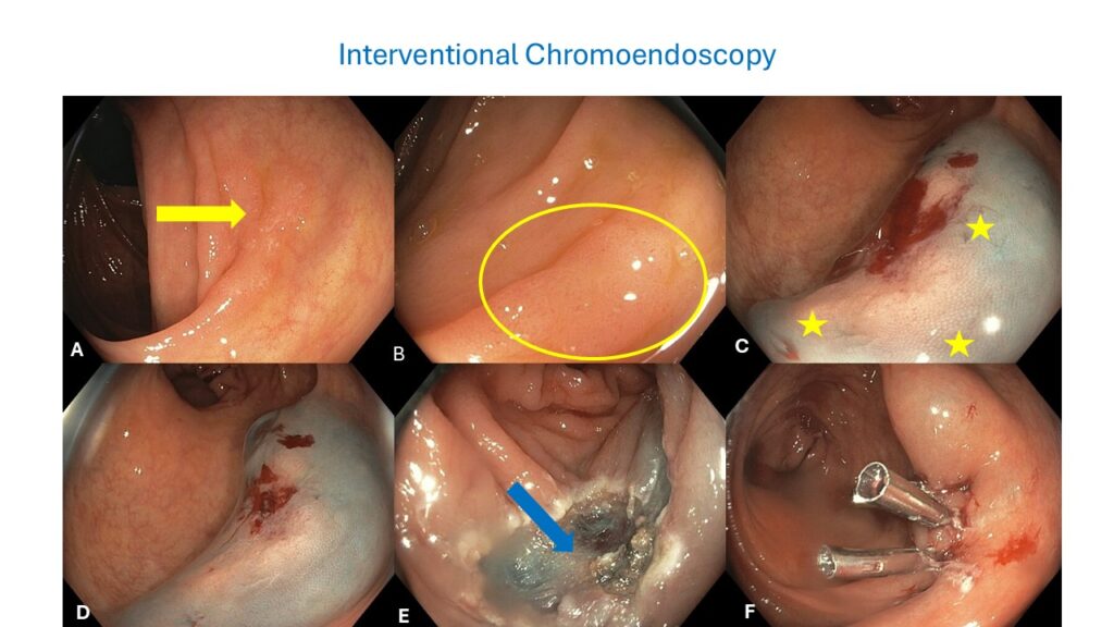
Figure 1 · Interventional chromoendoscopy

Endoscopic Findings
A flat lesion was identified in the colon (A, yellow arrow). Close-up examination revealed a small laterally spreading tumor measuring approximately 15 mm, classified as Paris 2a, with a Kudo pattern IIIS (B, yellow circle).
Endoscopic Technique
- The lesion was injected with Everlift, a submucosal lifting agent that includes a pre-mixed colored substance (C). This created a visible interface, with the neoplastic flat lesion appearing white against the blue submucosal layer (C and D).
- The presence of the blue color allowed for clear differentiation of the superficial lesion from deeper layers, such as the submucosa and muscularis propria (C, yellow stars).
- After resection, a white color corresponding to the muscularis propria was observed, indicating the depth of resection (E, blue arrow). The submucosa has a bluish color.
- Prophylactic clips were applied to close the post-resection defect (F).
Discussion
Interventional chromoendoscopy offers several advantages during endoscopic resection (EMR or ESD). First, it provides a color interface that delineates lesions that may be challenging to visualize under white light endoscopy, ensuring complete lesion characterization.
Second, the submucosal cushion created by the lifting agent provides a safety base for resection, minimizing the risk of perforation.
Third, it allows for clear visualization of the third space, including submucosal layers, providing a visual element to prevent complications. If the resection is carried out too deeply, the absence of blue color in the submucosa signals excessive depth, prompting prophylactic closure with clips to prevent immediate or delayed colon perforation.
Key Learning Points
01Interventional chromoendoscopy enhances visualization of subtle colorectal lesions.
02The technique creates a safety cushion, facilitating safe and complete endoscopic resection.
03The distinct color interface aids in assessing resection depth and preventing perforation.
04Prophylactic clip closure is crucial for deep resections to mitigate perforation risk.
References
- Parra-Blanco A, Gimeno-García AZ, Quintero E. Chromoendoscopy and new endoscopic imaging techniques. World J Gastroenterol. 2011;17(41):4541-4552.
- Kudo S, Hirota S, Nakajima T, et al. Colorectal tumors and pit pattern analysis. J Clin Gastroenterol. 1994;18(3):218-222.
- Tanaka S, Kashida H, Saito Y, et al. JGES guidelines for colorectal endoscopic submucosal dissection/endoscopic mucosal resection. Dig Endosc. 2020;32(1):154-180.
- Mönkemüller K, Wilcox CM. Interventional chromoendoscopy. Gastrointest Endosc. 2013;78:346-350.
For your teaching file
Save this case as a PDF
Opens the companion page on The Practicing Endoscopist.