Clinical Images
 Klaus Mönkemüller, MD, PhD, FASGE, FESGE, FJGES
Professor of Medicine, Virginia Tech Carilion School of Medicine, Roanoke, Virginia, USA
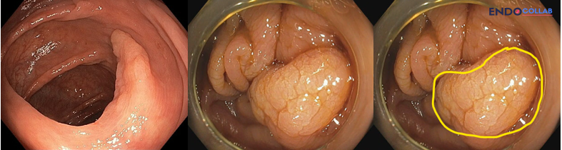
These images show acetic acid chromoendoscopy (2.5% acetic acid) for colon polyps. In the first example there is diffuse aceto-whitening. This is a serrated lesion.

In the second set of images there is no diffuse aceto-whitening, but there are areas of erythema (which represent low-grade and high-grade dysplasia). Histology showed a villous adenoma with low-grade dysplasia.

Acetic acid chromoendoscopy has tremendous use for defining serrated lesions. However, acetic acid may also be useful to define complex lateral spreading lesions, as shown in the second example.
Acetic acid chromoendoscopy is a specialized diagnostic technique that significantly improves the detection, delineation, and complete resection of serrated lesions (SLs), particularly sessile serrated lesions (SSLs), in the proximal colon. By temporarily altering mucosal proteins and dissolving surface mucus, acetic acid overcomes the traditional challenges of spotting these flat, pale, and easily missed precancerous polyps (1).
Get cases like this Saturday mornings. Free on The Practicing Endoscopist.
How Acetic Acid Chromoendoscopy Works
When a mild solution of acetic acid (typically 1.7% to 3%) is sprayed onto the colon lining via a catheter or syringe, it acts through a dual-action mechanism (1, 2):
01Mucolytic effect. The acid instantly dissolves the adherent mucus cap covering the polyp, exposing the true tissue surface beneath.
02Aceto-whitening (the "snowflake effect"). Acetic acid alters the cellular structure of glycoproteins and nuclear/cytoplasmic proteins. Because serrated lesions contain a high density of mucus-producing goblet cells, they react intensely to this process, turning a distinct white color (often referred to as the snowflake effect) (1, 2).
References
- Savóia de Oliveira FJ, Mota FL, Pereira Junior EMA, Loureiro JF, Navarro-Rodriguez T. Impact of acetic acid chromoendoscopy on detection of serrated lesions in the proximal colon compared with white-light endoscopy. Endosc Int Open. 2026;14:a28269621. doi:10.1055/a-2826-9621. PMID: 41970684.
- Yamamoto S, Varkey J, Bhandari P. Acetoelectronic chromoendoscopy for sessile serrated polyp. Gastrointest Endosc. 2021;93(1):267-268. doi:10.1016/j.gie.2020.07.008. PMID: 32679046.
For your teaching file
Save this case as a PDF
Opens the companion page on The Practicing Endoscopist.