Case Report

 Summer Stefanko, MD and Klaus Mönkemüller, MD, PhD, FASGE, FESGE, FJGES
Internal Medicine and Department of Gastroenterology, Virginia Tech Carilion School of Medicine, Roanoke, VA, USA
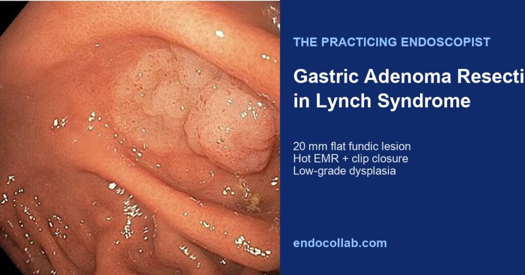
Figure 1 · Endoscopic findings


Experienced teaching points
Clinical Pearls
01Lynch syndrome predisposes patients to gastric adenocarcinoma, most often intestinal-type.
02ACG guidance supports considering EGD with gastric biopsy starting at ages 30 to 35, with H. pylori treatment if present.
03Surveillance EGDs can catch precancerous gastric adenomas early enough for endoscopic resection.
04Hot EMR with lifting agent and clip closure is an effective approach for selected flat fundic adenomas.
Get cases like this every other Saturday. Free on The Practicing Endoscopist.
Final Diagnosis
Gastric adenoma, intestinal type, with low-grade dysplasia, resected via endoscopic mucosal resection.
Clinical History
A 49-year-old man with Lynch syndrome presented for routine screening colonoscopy and esophagogastroduodenoscopy (EGD).
Endoscopic Findings
- EGD visualized a 20 mm flat polypoid lesion in the gastric fundus.
Endoscopic Technique
The lesion was resected with Eleview injection and hot endoscopic mucosal resection (EMR). The defect was closed with endoscopic clips. Histology showed gastric adenoma, intestinal type, with low-grade dysplasia. Testing was negative for Helicobacter pylori.
Discussion
Mechanism. Lynch syndrome is caused by autosomal dominant germline pathogenic variants in DNA mismatch repair genes. It is the most common hereditary cancer predisposition syndrome and raises lifetime risk for several cancers, especially colorectal (40 to 80%) and endometrial (40 to 60%) cancer. Gastric cancer risk ranges from about 0.2 to 13% depending on the gene mutation, and intestinal-type gastric adenocarcinoma is the usual histology.
What the scope shows. In this patient, a flat 20 mm fundic lesion was the actionable finding on surveillance EGD. Complete hot EMR with clip closure removed an intestinal-type adenoma with low-grade dysplasia before progression to invasive cancer.
Why it matters. ACG guidance recommends colonoscopy at least every two years in Lynch syndrome and supports considering EGD with gastric biopsy at ages 30 to 35, with H. pylori treatment if present and continued 3- to 5-year surveillance when family history includes gastric or duodenal cancer. This case shows how that surveillance pathway can catch and clear a precancerous gastric adenoma endoscopically.
References
- Kastrinos F, et al. The American College of Gastroenterology Clinical Guideline: Genetic Predisposition to Gastrointestinal Cancers. Am J Gastroenterol. 2021;116(11):2100-2122.
- Ladep NG, et al. Gastric cancer surveillance in Lynch syndrome: A systematic review. Fam Cancer. 2018;17(3):477-486.
- Pimentel-Nunes P, et al. Endoscopic mucosal resection and endoscopic submucosal dissection for gastric epithelial neoplasia: an ESGE Guideline. Endoscopy. 2015;47(9):829-856.
For your teaching file
Save this case as a PDF
Opens the companion page on The Practicing Endoscopist.