Technique Article
 Ali Safdar Khan, MD, MSPH and Klaus Mönkemüller, MD, PhD, FASGE (USA), FJGES (Japan), FESGE (Europe)
Professor of Medicine, Department of Gastroenterology, Ameos Klinikum Halberstadt, Germany and Carilion Memorial Hospital, Virginia Tech Carilion School of Medicine, Roanoke, USA
Experienced teaching points
Clinical Pearls
01Direct cholangioscopy using ultraslim endoscopes may fail in up to 15% of cases, mainly due to cannulation failure from a stenotic bile duct or difficult scope position.
02Placement of a fully covered self-expanding metal stent (fcSEMS) in the bile duct can facilitate direct biliary access by providing a channel for the ultraslim scope.
03Stent-assisted direct cholangioscopy is a novel technique that enables both diagnostic and therapeutic interventions of the biliary tract when conventional direct cholangioscopy is not feasible.
04This approach allowed successful removal of residual stones using direct observation cholangioscopy (DOC) with basket through the fcSEMS.
Get cases like this every other Saturday. Free on The Practicing Endoscopist.
Clinical History
Direct cholangioscopy using ultra slims endoscopes (i.e. pediatric gastroscopes or transnasal gastroscopes) enables diagnostic and therapeutic interventions of the biliary tract. However, DC is not always feasible with failure rates up to 15%, mainly due to cannulation failure. Entering the bile duct may be hampered by a stenotic bile duct and difficult scope position. Herein we present the novel concept of stent-assisted direct cholangioscopy.
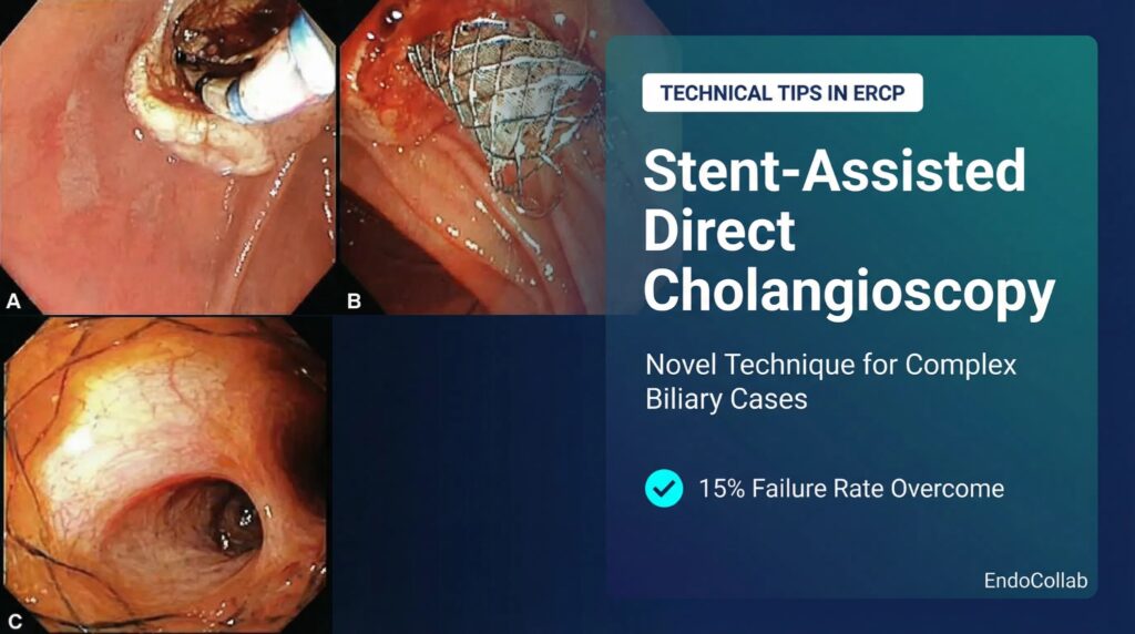
A 76-year-old female patient was referred to our institution with obstructive jaundice due to complex biliary tract lithiasis, which could not be removed during the initial ERCP. At our hospital cholangiography revealed a very dilated, tortuous bile duct with filling defects in the common hepatic and right intrahepatic duct. At the referring hospital the stone could not be retrieved, and thus dual biliary stent insertion (7fr 15cm and 10Fr 5cm) was done. At our hospital ERCP performed 12 weeks later revealed a narrowed and fibrosed papilla. After removing the plastic stents and performing biliary sphincterotomy (panel A) an extraction balloon was used to sweep significant amount of debris out of the severely dilated bile ducts. Given the previous concern for stones and the severely dilated biliary tree, a fully covered metal stent (10Fr 8cm) (Cook Medical, USA) was placed in the bile duct (B). A 4.9 mm ultraslim scope was passed nasally into the stomach and duodenum and then the bile duct was cannulated through the fcSEMS, thus achieving a direct stent-assisted cholangioscopy (C). The remaining stones were removed using DOC with basket. No stones were left behind intraductally (C).
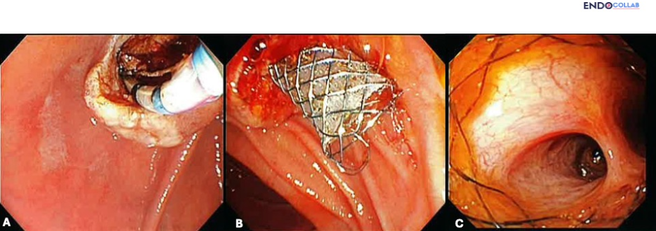
This case shows how inserting a fully covered self-expanding metal stent allowed for direct biliary access using an ultraslim transnasal gastroscope, thus performing direct cholangioscopy.
For your teaching file
Save this case as a PDF
Opens the companion page on The Practicing Endoscopist.