Case Report
 Klaus Mönkemüller, MD, PhD, FASGE, FESGE, FJGES
Professor of Medicine, Department of Gastroenterology, Carilion Memorial Hospital, Virginia Tech Carilion School of Medicine, Roanoke, USA
Experienced teaching points
Clinical Pearls
01Acetic acid chromoendoscopy enhances the detection of dysplasia in Barrett esophagus by highlighting areas with altered cellular pathology.
02A 2.5% acetic acid solution is optimal for Barrett esophagus given concerns for cell damage and accuracy with higher concentrations.
03The phenomenon of "loss of aceto-whitening" indicates dysplastic or cancerous changes due to differential cellular responses to acetic acid.
04Targeted biopsies of areas with loss of aceto-whitening improve diagnostic yield for high-grade dysplasia and early cancer in Barrett esophagus.
Get cases like this every other Saturday. Free on The Practicing Endoscopist.
Clinical History
Two patients, a 54-year-old male with a 10-year history of Barrett esophagus and a 75-year-old male with a 20-year history of Barrett esophagus, presented for routine surveillance endoscopy. In both cases, the primary goal was dysplasia detection using acetic acid chromoendoscopy.

Endoscopic Findings
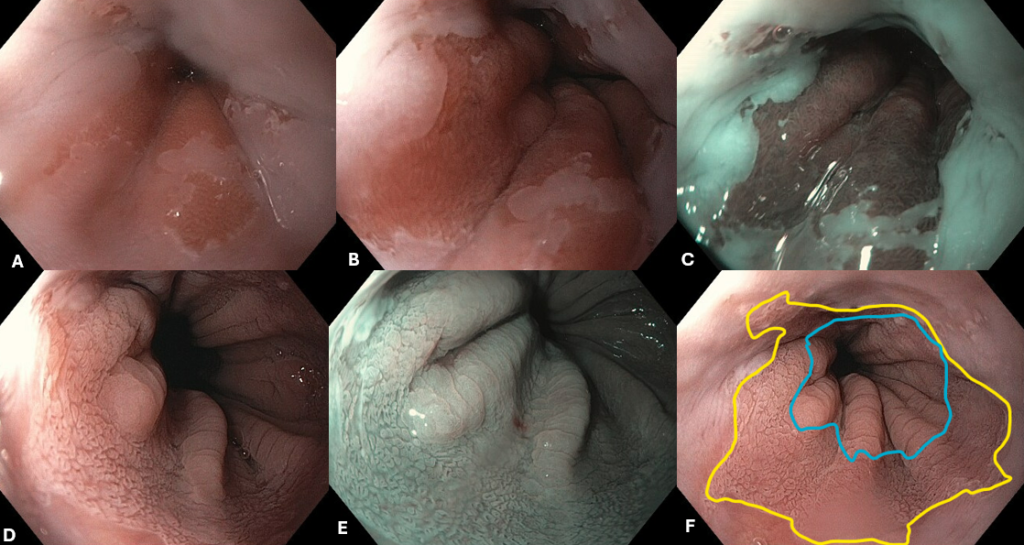
In the first patient, no suspicious mucosal features were identified within the Barrett segment, which extended 15 mm proximal to the gastric folds.
In the second patient, after approximately 10 minutes of acetic acid application, distinct areas showing a loss of aceto-whitening (appearing red against the surrounding white coagulated mucosa) were observed. These specific red areas were targeted for biopsy.
Endoscopic Technique
Standard upper endoscopy was performed first. Acetic acid chromoendoscopy was executed using a 2.5% solution. High concentrations (such as 5%) were avoided due to the risk of cellular damage and reduced diagnostic accuracy from excessive whitening, which can mask suspicious areas.
Acetic acid breaks disulfide bonds in the mucin glycoproteins of the cylindrical epithelial cells, permitting the acid to enter the cytoplasm. The weak acid causes reversible denaturation and coagulation of cytoplasmic proteins, including structural cytokeratins. This coagulated protein matrix scatters endoscopic light, making the tissue appear white and masking the vascular pattern.
In dysplastic or cancerous cells, the cytoplasmic proteins have a reduced capacity to retain the acid due to abnormal, overcrowded nuclei. The whitening effect dissipates rapidly, resulting in a visible redness (loss of aceto-whitening). Targeted biopsies were obtained from these red zones.
Discussion
Acetic acid chromoendoscopy is a low-cost, valuable technique for detecting dysplasia in Barrett esophagus. The method relies on the differential cellular response. Normal Barrett epithelium whitens and retains the effect for several minutes. In contrast, dysplastic cells show transient whitening followed by rapid clearance, appearing red. This biological response allows the endoscopist to transition from random biopsy protocols to targeted biopsies, which significantly improves diagnostic yield. In the second patient, biopsies from the red zones showing loss of aceto-whitening confirmed high-grade dysplasia. For other cases involving dysplasia and mucosal surveillance, see our guide on pedunculated polyp resection and surveillance.
References
- Pohl H, Rösch T, Wallace MB. Acetic acid chromoendoscopy for Barrett’s esophagus. Endoscopy. 2008;40(7):594-599.
- Curvers WL, Alvarez Herrero L, Fockens P, et al. Mucosal morphology in Barrett’s esophagus: correlation between magnification endoscopy and histology. Clin Gastroenterol Hepatol. 2008;6(2):167-173.
- Singh R, Sharma S, Rastogi A, et al. Acetic Acid Chromoendoscopy for Early Detection of Dysplasia in Barrett’s Esophagus: A Meta-Analysis. Dig Dis Sci. 2018;63(1):1-10.
For your teaching file
Save this case as a PDF
Opens the companion page on The Practicing Endoscopist.