
Diagnosis at First Sight: Gastric Adenoma
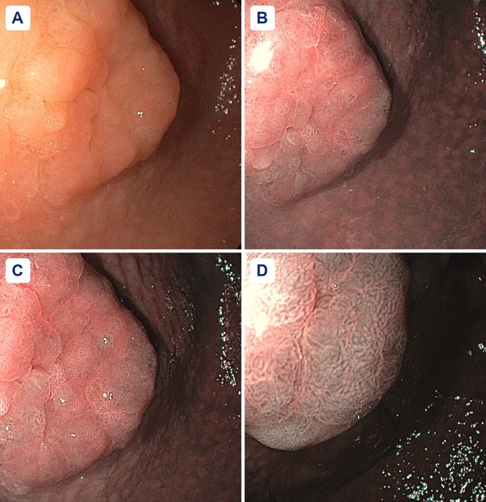
A raised 2 cm lesion near the incisura, found on an EGD for reflux. White light, narrow-band imaging, and why histology remains necessary for diagnosis and grading.
Vanek Tumor of the Stomach
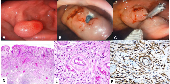
A sessile 18 to 20 mm antral polyp in a 50-year-old woman. Endoscopic mucosal resection with clip closure, and immunohistochemistry (CD34 positive, CD117 and DOG1 negative) that ruled out GIST.
Acetic Acid (Vinegar) Chromoendoscopy for Colon Polyps
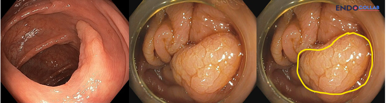
Diffuse aceto-whitening marks a serrated lesion. Focal erythema without whitening pointed to dysplasia in a villous adenoma. How 2.5% acetic acid chromoendoscopy works in the colon.
When to Perform Endoscopy or Radiological Tests in Patients with Unresponsive Celiac Disease
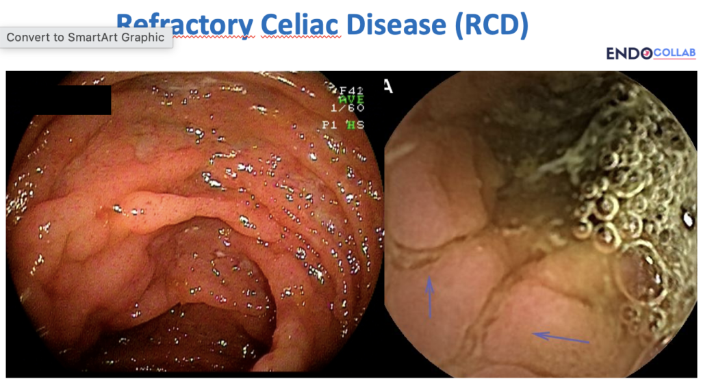
Persistent or worsening celiac symptoms warrant workup for refractory disease, EATL, and adenocarcinoma, plus key endoscopic and radiologic warning signs.
An Exophytic Ileocecal Mass: Diffuse Large B-Cell Lymphoma Diagnosed by Colonoscopic Biopsy
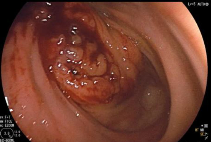
Exophytic ileocecal-valve mass on colonoscopy; biopsy confirmed germinal-center DLBCL. Keep lymphoma on the differential; do not treat as adenocarcinoma by default.
Endoscopic Management of a Duodenal Dieulafoy’s Lesion
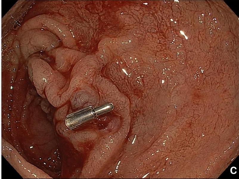
Spurting D2 Dieulafoy in a patient with hematochezia: protruding vessel, no ulcer crater. One through-the-scope clip stopped the bleed immediately.
Plummer-Vinson Syndrome with Celiac Disease and Esophageal Varices
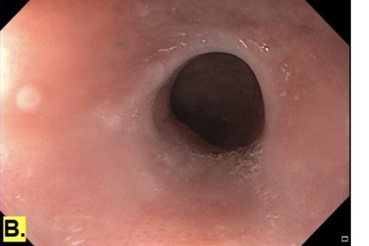
Plummer-Vinson with iron-deficiency anemia: proximal esophageal webs needing dilation, plus celiac and varices. Treat the web, then hunt the shared iron-loss driver.
How Would You Remove This Pedunculated Polyp (Paris 0-Ip, Kudo IIIL)?
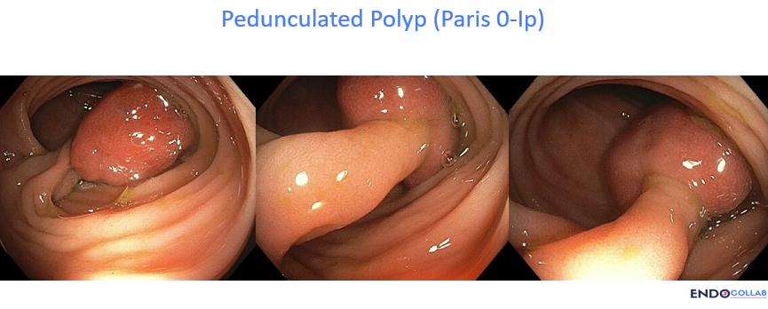
A large sigmoid 0-Ip polyp in a patient on Eliquis. Ten ways to resect it, and why prophylactic hemostasis comes before the cut.
Post-Prandial Thoracic Pain and Pre-Syncopal Symptoms: Watch Out for Roemheld Syndrome
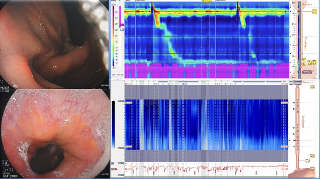
A 26-year-old woman with postprandial chest pain and palpitations after a negative cardiac workup. Large hiatal hernia, reflux esophagitis, ineffective esophageal motility, and pathologic reflux: Roemheld syndrome.
Portal Vein-Duodenal Fistula After Sequential Abdominal Surgery
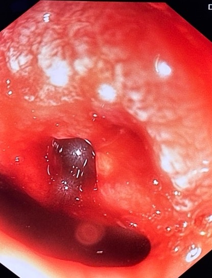
A 57-year-old man developed massive hematemesis after laparoscopic cholecystectomy and laparotomy for perforated appendicitis. Second-look EGD found a fistulous orifice in the duodenal bulb. CT showed a right portal vein pseudoaneurysm. Transcatheter coil embolization closed it.
Ménétrier Disease: A Diagnostic Challenge of Enlarged Gastric Folds
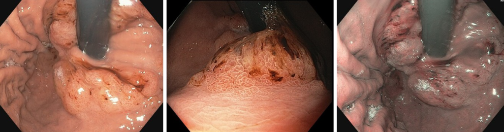
A 76-year-old man with anemia had giant gastric folds that did not flatten with air. EUS showed intact muscularis propria; deep biopsies confirmed Ménétrier disease.
Gastric Adenoma Resection in a Patient with Lynch Syndrome
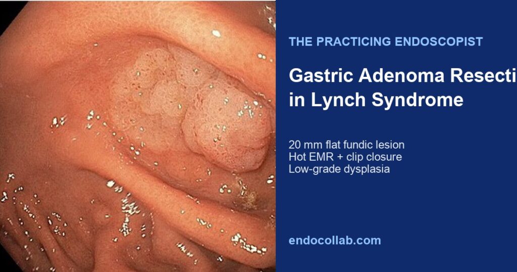
Surveillance EGD in Lynch syndrome found a 20 mm flat fundic adenoma. Hot EMR resected intestinal-type adenoma with low-grade dysplasia.